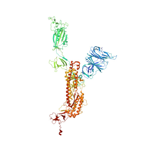
7BNO

Open conformation of D614G SARS-CoV-2 spike with 2 Erect RBDs


Experimental Data Snapshot

  • Method: ELECTRON MICROSCOPY
  • Resolution: 4.20 Å
  • Aggregation State: PARTICLE 
  • Reconstruction Method: SINGLE PARTICLE 

wwPDB Validation   3D Report Full Report


This is version 1.3 of the entry. See complete history


Literature

The effect of the D614G substitution on the structure of the spike glycoprotein of SARS-CoV-2.

Benton, D.J.Wrobel, A.G.Roustan, C.Borg, A.Xu, P.Martin, S.R.Rosenthal, P.B.Skehel, J.J.Gamblin, S.J.

(2021) Proc Natl Acad Sci U S A 118

  • DOI: https://doi.org/10.1073/pnas.2022586118
  • Primary Citation of Related Structures:  
    7BNM, 7BNN, 7BNO

  • PubMed Abstract: 

    The majority of currently circulating severe acute respiratory syndrome coronavirus 2 (SARS-CoV-2) viruses have mutant spike glycoproteins that contain the D614G substitution. Several studies have suggested that spikes with this substitution are associated with higher virus infectivity. We use cryo-electron microscopy to compare G614 and D614 spikes and show that the G614 mutant spike adopts a range of more open conformations that may facilitate binding to the SARS-CoV-2 receptor, ACE2, and the subsequent structural rearrangements required for viral membrane fusion.


  • Organizational Affiliation

    Structural Biology of Disease Processes Laboratory, Francis Crick Institute, London NW1 1AT, United Kingdom; Donald.Benton@crick.ac.uk John.Skehel@crick.ac.uk Antoni.Wrobel@crick.ac.uk Steve.Gamblin@crick.ac.uk.


Macromolecules
Find similar proteins by:  (by identity cutoff)  |  3D Structure
Entity ID: 1
MoleculeChains Sequence LengthOrganismDetailsImage
Spike glycoprotein
A, B, C
1,177Severe acute respiratory syndrome coronavirus 2Mutation(s): 0 
Gene Names: S2
UniProt
Find proteins for P0DTC2 (Severe acute respiratory syndrome coronavirus 2)
Explore P0DTC2 
Go to UniProtKB:  P0DTC2
Entity Groups  
Sequence Clusters30% Identity50% Identity70% Identity90% Identity95% Identity100% Identity
UniProt GroupP0DTC2
Glycosylation
Glycosylation Sites: 12Go to GlyGen: P0DTC2-1
Sequence Annotations
Expand
  • Reference Sequence
Oligosaccharides

Help

Entity ID: 2
MoleculeChains Length2D Diagram Glycosylation3D Interactions
2-acetamido-2-deoxy-beta-D-glucopyranose-(1-4)-2-acetamido-2-deoxy-beta-D-glucopyranose
D
2N-Glycosylation
Glycosylation Resources
GlyTouCan:  G42666HT
GlyCosmos:  G42666HT
GlyGen:  G42666HT
Small Molecules
Ligands 1 Unique
IDChains Name / Formula / InChI Key2D Diagram3D Interactions
NAG
Query on NAG

Download Ideal Coordinates CCD File 
AA [auth C]
BA [auth C]
CA [auth C]
DA [auth C]
E [auth A]
AA [auth C],
BA [auth C],
CA [auth C],
DA [auth C],
E [auth A],
EA [auth C],
F [auth A],
FA [auth C],
G [auth A],
GA [auth C],
H [auth A],
HA [auth C],
I [auth A],
IA [auth C],
J [auth A],
K [auth A],
L [auth A],
M [auth A],
N [auth A],
O [auth A],
P [auth B],
Q [auth B],
R [auth B],
S [auth B],
T [auth B],
U [auth B],
V [auth B],
W [auth B],
X [auth B],
Y [auth B],
Z [auth C]
2-acetamido-2-deoxy-beta-D-glucopyranose
C8 H15 N O6
OVRNDRQMDRJTHS-FMDGEEDCSA-N
Experimental Data & Validation

Experimental Data

  • Method: ELECTRON MICROSCOPY
  • Resolution: 4.20 Å
  • Aggregation State: PARTICLE 
  • Reconstruction Method: SINGLE PARTICLE 

Structure Validation

View Full Validation Report


Entry History & Funding Information

Deposition Data


Funding OrganizationLocationGrant Number
The Francis Crick InstituteUnited KingdomFC001078
The Francis Crick InstituteUnited KingdomFC001143

Revision History  (Full details and data files)

  • Version 1.0: 2021-02-03
    Type: Initial release
  • Version 1.1: 2021-02-24
    Changes: Database references
  • Version 1.2: 2021-03-10
    Changes: Database references, Source and taxonomy, Structure summary
  • Version 1.3: 2024-10-23
    Changes: Data collection, Database references, Structure summary