7DRB | pdb_00007drb

Crystal structure of plant receptor like protein RXEG1 with xyloglucanase XEG1


Experimental Data Snapshot

  • Method: X-RAY DIFFRACTION
  • Resolution: 3.30 Å
  • R-Value Free: 
    0.284 (Depositor), 0.290 (DCC) 
  • R-Value Work: 
    0.227 (Depositor), 0.240 (DCC) 
  • R-Value Observed: 
    0.230 (Depositor) 

Starting Model: experimental
View more details

wwPDB Validation   3D Report Full Report


Ligand Structure Quality Assessment 

Created with Raphaël 2.3.0Worse 01 BetterLigand structure goodness of fit to experimental dataBest fitted NAGClick on this verticalbar to view details

This is version 1.3 of the entry. See complete history


Literature

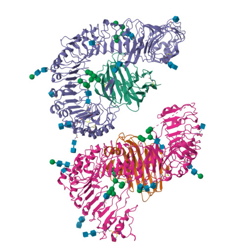
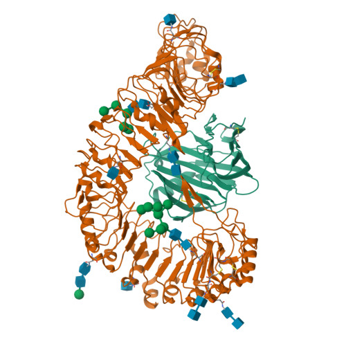
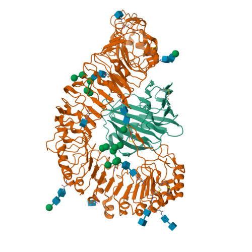
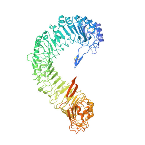
Plant receptor-like protein activation by a microbial glycoside hydrolase.

Sun, Y.Wang, Y.Zhang, X.Chen, Z.Xia, Y.Wang, L.Sun, Y.Zhang, M.Xiao, Y.Han, Z.Wang, Y.Chai, J.

(2022) Nature 610: 335-342

  • DOI: https://doi.org/10.1038/s41586-022-05214-x
  • Primary Citation of Related Structures:  
    7DRB, 7DRC, 7W3T, 7W3V, 7W3X

  • PubMed Abstract: 

    Plants rely on cell-surface-localized pattern recognition receptors to detect pathogen- or host-derived danger signals and trigger an immune response 1-6 . Receptor-like proteins (RLPs) with a leucine-rich repeat (LRR) ectodomain constitute a subgroup of pattern recognition receptors and play a critical role in plant immunity 1-3 . Mechanisms underlying ligand recognition and activation of LRR-RLPs remain elusive. Here we report a crystal structure of the LRR-RLP RXEG1 from Nicotiana benthamiana that recognizes XEG1 xyloglucanase from the pathogen Phytophthora sojae. The structure reveals that specific XEG1 recognition is predominantly mediated by an amino-terminal and a carboxy-terminal loop-out region (RXEG1(ID)) of RXEG1. The two loops bind to the active-site groove of XEG1, inhibiting its enzymatic activity and suppressing Phytophthora infection of N. benthamiana. Binding of XEG1 promotes association of RXEG1(LRR) with the LRR-type co-receptor BAK1 through RXEG1(ID) and the last four conserved LRRs to trigger RXEG1-mediated immune responses. Comparison of the structures of apo-RXEG1(LRR), XEG1-RXEG1(LRR) and XEG1-BAK1-RXEG1(LRR) shows that binding of XEG1 induces conformational changes in the N-terminal region of RXEG1(ID) and enhances structural flexibility of the BAK1-associating regions of RXEG1(LRR). These changes allow fold switching of RXEG1(ID) for recruitment of BAK1(LRR). Our data reveal a conserved mechanism of ligand-induced heterodimerization of an LRR-RLP with BAK1 and suggest a dual function for the LRR-RLP in plant immunity.


  • Organizational Affiliation

    Tsinghua-Peking Center for Life Sciences, Beijing Advanced Innovation Center for Structural Biology, Center for Plant Biology, School of Life Sciences, Tsinghua University, Beijing, China.


Macromolecules
Find similar proteins by:  (by identity cutoff)  |  3D Structure
Entity ID: 1
MoleculeChains Sequence LengthOrganismDetailsImage
Cell 12A endoglucanaseA,
C [auth B]
241Phytophthora sojaeMutation(s): 0 
Gene Names: EGL12-G
UniProt
Find proteins for Q30BZ2 (Phytophthora sojae)
Explore Q30BZ2 
Go to UniProtKB:  Q30BZ2
Entity Groups  
Sequence Clusters30% Identity50% Identity70% Identity90% Identity95% Identity100% Identity
UniProt GroupQ30BZ2
Sequence Annotations
Expand
  • Reference Sequence
Find similar proteins by:  (by identity cutoff)  |  3D Structure
Entity ID: 2
MoleculeChains Sequence LengthOrganismDetailsImage
Membrane-localized LRR receptor-like proteinB [auth C],
D
934Nicotiana benthamianaMutation(s): 0 
Gene Names: RXEG1
UniProt
Find proteins for A0A2I8B6R1 (Nicotiana benthamiana)
Explore A0A2I8B6R1 
Go to UniProtKB:  A0A2I8B6R1
Entity Groups  
Sequence Clusters30% Identity50% Identity70% Identity90% Identity95% Identity100% Identity
UniProt GroupA0A2I8B6R1
Glycosylation
Glycosylation Sites: 10
Sequence Annotations
Expand
  • Reference Sequence
Oligosaccharides

Help

Entity ID: 3
MoleculeChains Length2D Diagram Glycosylation3D Interactions
alpha-D-mannopyranose-(1-2)-alpha-D-mannopyranose-(1-3)-[alpha-D-mannopyranose-(1-2)-alpha-D-mannopyranose-(1-6)]alpha-D-mannopyranose-(1-6)-[alpha-D-mannopyranose-(1-2)-alpha-D-mannopyranose-(1-3)]beta-D-mannopyranose-(1-4)-2-acetamido-2-deoxy-beta-D-glucopyranose-(1-4)-2-acetamido-2-deoxy-beta-D-glucopyranose
E, L
10N-Glycosylation
Glycosylation Resources
GlyTouCan:  G89864BN
GlyCosmos:  G89864BN
GlyGen:  G89864BN
Entity ID: 4
MoleculeChains Length2D Diagram Glycosylation3D Interactions
2-acetamido-2-deoxy-beta-D-glucopyranose-(1-4)-2-acetamido-2-deoxy-beta-D-glucopyranose
F, G, J, K, M
2N-Glycosylation
Glycosylation Resources
GlyTouCan:  G42666HT
GlyCosmos:  G42666HT
GlyGen:  G42666HT
Entity ID: 5
MoleculeChains Length2D Diagram Glycosylation3D Interactions
beta-D-mannopyranose-(1-4)-2-acetamido-2-deoxy-beta-D-glucopyranose-(1-4)-2-acetamido-2-deoxy-beta-D-glucopyranose
H, N, O, R
3N-Glycosylation
Glycosylation Resources
GlyTouCan:  G15407YE
GlyCosmos:  G15407YE
GlyGen:  G15407YE
Entity ID: 6
MoleculeChains Length2D Diagram Glycosylation3D Interactions
alpha-D-mannopyranose-(1-2)-alpha-D-mannopyranose-(1-3)-[alpha-D-mannopyranose-(1-6)-alpha-D-mannopyranose-(1-6)]beta-D-mannopyranose-(1-4)-2-acetamido-2-deoxy-beta-D-glucopyranose-(1-4)-2-acetamido-2-deoxy-beta-D-glucopyranose
I, P
7N-Glycosylation
Glycosylation Resources
GlyTouCan:  G23799GS
GlyCosmos:  G23799GS
GlyGen:  G23799GS
Experimental Data & Validation

Experimental Data

  • Method: X-RAY DIFFRACTION
  • Resolution: 3.30 Å
  • R-Value Free:  0.284 (Depositor), 0.290 (DCC) 
  • R-Value Work:  0.227 (Depositor), 0.240 (DCC) 
  • R-Value Observed: 0.230 (Depositor) 
Space Group: P 1 21 1
Unit Cell:
Length ( Å )Angle ( ˚ )
a = 103.888α = 90
b = 92.844β = 107.91
c = 154.526γ = 90
Software Package:
Software NamePurpose
PHENIXrefinement
HKL-2000data scaling
PDB_EXTRACTdata extraction
HKL-2000data reduction
PHENIXphasing

Structure Validation

View Full Validation Report


Ligand Structure Quality Assessment 

Created with Raphaël 2.3.0Worse 01 BetterLigand structure goodness of fit to experimental dataBest fitted NAGClick on this verticalbar to view details

Entry History & Funding Information

Deposition Data


Funding OrganizationLocationGrant Number
National Natural Science Foundation of China (NSFC)China--
Alexander von Humboldt FoundationGermany--

Revision History  (Full details and data files)

  • Version 1.0: 2022-06-22
    Type: Initial release
  • Version 1.1: 2022-10-26
    Changes: Database references
  • Version 1.2: 2023-11-29
    Changes: Data collection, Refinement description
  • Version 1.3: 2024-10-23
    Changes: Structure summary