7E19

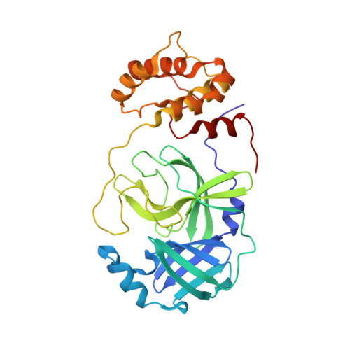
Crystal structure of SAR-CoV-2 3CL protease complex with inhibitor SH-5


Experimental Data Snapshot

  • Method: X-RAY DIFFRACTION
  • Resolution: 2.15 Å
  • R-Value Free: 0.235 
  • R-Value Work: 0.191 
  • R-Value Observed: 0.193 

Starting Model: experimental
View more details

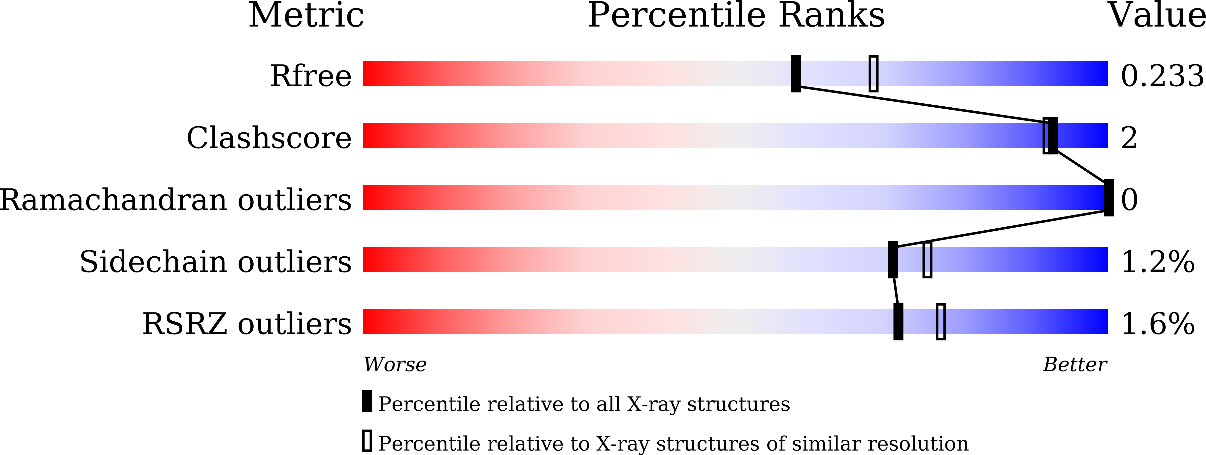
wwPDB Validation   3D Report Full Report


Ligand Structure Quality Assessment 


This is version 1.3 of the entry. See complete history


Literature

3CL Protease Inhibitors with an Electrophilic Arylketone Moiety as Anti-SARS-CoV-2 Agents.

Konno, S.Kobayashi, K.Senda, M.Funai, Y.Seki, Y.Tamai, I.Schakel, L.Sakata, K.Pillaiyar, T.Taguchi, A.Taniguchi, A.Gutschow, M.Muller, C.E.Takeuchi, K.Hirohama, M.Kawaguchi, A.Kojima, M.Senda, T.Shirasaka, Y.Kamitani, W.Hayashi, Y.

(2022) J Med Chem 65: 2926-2939

  • DOI: https://doi.org/10.1021/acs.jmedchem.1c00665
  • Primary Citation of Related Structures:  
    7E18, 7E19

  • PubMed Abstract: 

    The novel coronavirus, SARS-CoV-2, has been identified as the causative agent for the current coronavirus disease (COVID-19) pandemic. 3CL protease (3CL pro ) plays a pivotal role in the processing of viral polyproteins. We report peptidomimetic compounds with a unique benzothiazolyl ketone as a warhead group, which display potent activity against SARS-CoV-2 3CL pro . The most potent inhibitor YH-53 can strongly block the SARS-CoV-2 replication. X-ray structural analysis revealed that YH-53 establishes multiple hydrogen bond interactions with backbone amino acids and a covalent bond with the active site of 3CL pro . Further results from computational and experimental studies, including an in vitro absorption, distribution, metabolism, and excretion profile, in vivo pharmacokinetics, and metabolic analysis of YH-53 suggest that it has a high potential as a lead candidate to compete with COVID-19.


  • Organizational Affiliation

    School of Pharmacy, Department of Medicinal Chemistry, Tokyo University of Pharmacy and Life Sciences, Hachioji, Tokyo, 192-0392, Japan.


Macromolecules
Find similar proteins by:  (by identity cutoff)  |  3D Structure
Entity ID: 1
MoleculeChains Sequence LengthOrganismDetailsImage
3C-like proteinase306Severe acute respiratory syndrome coronavirus 2Mutation(s): 0 
EC: 3.4.22.69
UniProt
Find proteins for P0DTD1 (Severe acute respiratory syndrome coronavirus 2)
Explore P0DTD1 
Go to UniProtKB:  P0DTD1
Entity Groups  
Sequence Clusters30% Identity50% Identity70% Identity90% Identity95% Identity100% Identity
UniProt GroupP0DTD1
Sequence Annotations
Expand
  • Reference Sequence
Small Molecules
Ligands 1 Unique
IDChains Name / Formula / InChI Key2D Diagram3D Interactions
HUO (Subject of Investigation/LOI)
Query on HUO

Download Ideal Coordinates CCD File 
B [auth A](phenylmethyl) N-[(2S)-1-[[(2S)-1-[[(2S)-1-(1,3-benzothiazol-2-yl)-1-oxidanylidene-3-[(3S)-2-oxidanylidenepyrrolidin-3-yl]propan-2-yl]amino]-4-methyl-1-oxidanylidene-pentan-2-yl]amino]-3-methyl-1-oxidanylidene-butan-2-yl]carbamate
C33 H41 N5 O6 S
KSCRVOKQPYZBHZ-IXPOFIJOSA-N
Experimental Data & Validation

Experimental Data

  • Method: X-RAY DIFFRACTION
  • Resolution: 2.15 Å
  • R-Value Free: 0.235 
  • R-Value Work: 0.191 
  • R-Value Observed: 0.193 
  • Space Group: P 21 21 2
Unit Cell:
Length ( Å )Angle ( ˚ )
a = 45.111α = 90
b = 63.286β = 90
c = 106.697γ = 90
Software Package:
Software NamePurpose
PHENIXrefinement
XDSdata reduction
XSCALEdata scaling
MOLREPphasing
Cootmodel building

Structure Validation

View Full Validation Report


Ligand Structure Quality Assessment 


Entry History & Funding Information

Deposition Data


Funding OrganizationLocationGrant Number
Japan Agency for Medical Research and Development (AMED)JapanJP20am0101001

Revision History  (Full details and data files)

  • Version 1.0: 2021-06-23
    Type: Initial release
  • Version 1.1: 2021-08-11
    Changes: Database references
  • Version 1.2: 2022-03-09
    Changes: Database references
  • Version 1.3: 2023-11-29
    Changes: Data collection, Refinement description