7FH0

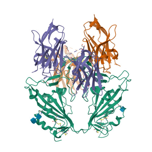
Crystallographic structure of two neutralizing nanobodies in complex with SARS-CoV-2 spike receptor-binding Domain (RBD)


Experimental Data Snapshot

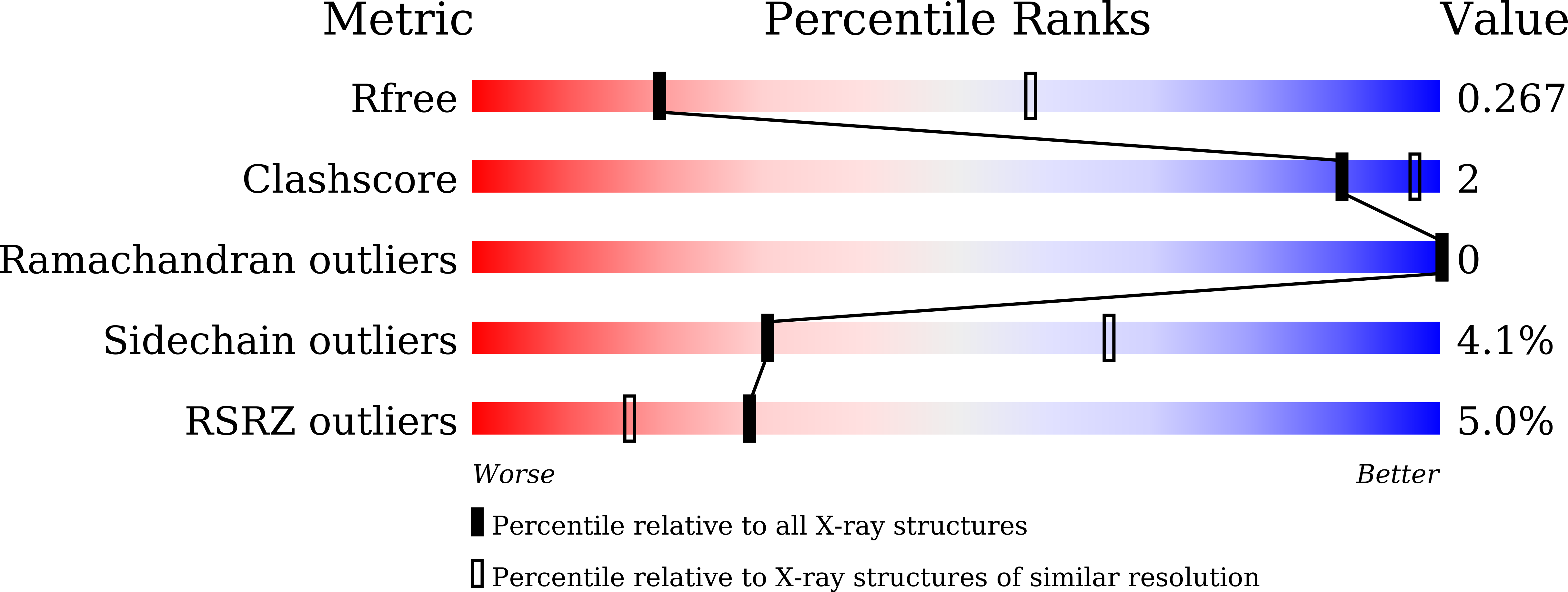
  • Method: X-RAY DIFFRACTION
  • Resolution: 3.20 Å
  • R-Value Free: 0.263 
  • R-Value Work: 0.207 
  • R-Value Observed: 0.210 

Starting Models: experimental
View more details

wwPDB Validation   3D Report Full Report


Ligand Structure Quality Assessment 


This is version 1.4 of the entry. See complete history


Literature

Hetero-bivalent nanobodies provide broad-spectrum protection against SARS-CoV-2 variants of concern including Omicron.

Ma, H.Zhang, X.Zheng, P.Dube, P.H.Zeng, W.Chen, S.Cheng, Q.Yang, Y.Wu, Y.Zhou, J.Hu, X.Xiang, Y.Zhang, H.Chiu, S.Jin, T.

(2022) Cell Res 32: 831-842

  • DOI: https://doi.org/10.1038/s41422-022-00700-3
  • Primary Citation of Related Structures:  
    7FH0, 7VOA

  • PubMed Abstract: 

    SARS-CoV-2 variants with adaptive mutations have continued to emerge, causing fresh waves of infection even amongst vaccinated population. The development of broad-spectrum antivirals is thus urgently needed. We previously developed two hetero-bivalent nanobodies (Nbs), aRBD-2-5 and aRBD-2-7, with potent neutralization activity against the wild-type (WT) Wuhan isolated SARS-CoV-2, by fusing aRBD-2 with aRBD-5 and aRBD-7, respectively. Here, we resolved the crystal structures of these Nbs in complex with the receptor-binding domain (RBD) of the spike protein, and found that aRBD-2 contacts with highly-conserved RBD residues and retains binding to the RBD of the Alpha, Beta, Gamma, Delta, Delta plus, Kappa, Lambda, Omicron BA.1, and BA.2 variants. In contrast, aRBD-5 and aRBD-7 bind to less-conserved RBD epitopes non-overlapping with the epitope of aRBD-2, and do not show apparent binding to the RBD of some variants. However, when fused with aRBD-2, they effectively enhance the overall binding affinity. Consistently, aRBD-2-5-Fc and aRBD-2-7-Fc potently neutralized all of the tested authentic or pseudotyped viruses, including WT, Alpha, Beta, Gamma, Delta, and Omicron BA.1, BA.1.1 and BA.2. Furthermore, aRBD-2-5-Fc provided prophylactic protection against the WT and mouse-adapted SARS-CoV-2 in mice, and conferred protection against the Omicron BA.1 variant in hamsters prophylactically and therapeutically, indicating that aRBD-2-5-Fc could potentially benefit the prevention and treatment of COVID-19 caused by the emerging variants of concern. Our strategy provides new solutions in the development of broad-spectrum therapeutic antibodies for COVID-19.


  • Organizational Affiliation

    Department of Pulmonary and Critical Care Medicine, The First Affiliated Hospital of USTC, Division of Life Sciences and Medicine, University of Science and Technology of China, Hefei, Anhui, China.


Macromolecules
Find similar proteins by:  (by identity cutoff)  |  3D Structure
Entity ID: 1
MoleculeChains Sequence LengthOrganismDetailsImage
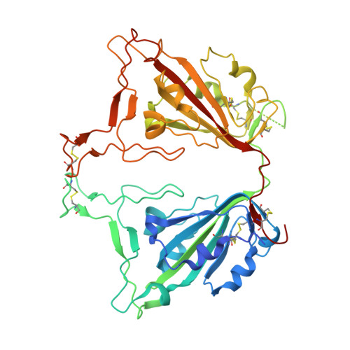
Spike protein S1,Spike protein S1438Severe acute respiratory syndrome coronavirus 2Mutation(s): 0 
Gene Names: S2
UniProt
Find proteins for P0DTC2 (Severe acute respiratory syndrome coronavirus 2)
Explore P0DTC2 
Go to UniProtKB:  P0DTC2
Entity Groups  
Sequence Clusters30% Identity50% Identity70% Identity90% Identity95% Identity100% Identity
UniProt GroupP0DTC2
Glycosylation
Glycosylation Sites: 2Go to GlyGen: P0DTC2-1
Sequence Annotations
Expand
  • Reference Sequence
Find similar proteins by:  (by identity cutoff)  |  3D Structure
Entity ID: 2
MoleculeChains Sequence LengthOrganismDetailsImage
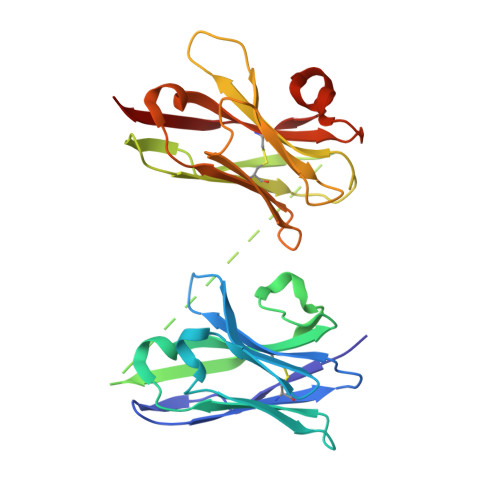
nanobodies aRBD-2-7
B, C
261Vicugna pacosMutation(s): 0 
Entity Groups  
Sequence Clusters30% Identity50% Identity70% Identity90% Identity95% Identity100% Identity
Sequence Annotations
Expand
  • Reference Sequence
Oligosaccharides

Help

Entity ID: 3
MoleculeChains Length2D Diagram Glycosylation3D Interactions
2-acetamido-2-deoxy-beta-D-glucopyranose-(1-4)-2-acetamido-2-deoxy-beta-D-glucopyranose
D
2N-Glycosylation
Glycosylation Resources
GlyTouCan:  G42666HT
GlyCosmos:  G42666HT
GlyGen:  G42666HT
Small Molecules
Experimental Data & Validation

Experimental Data

  • Method: X-RAY DIFFRACTION
  • Resolution: 3.20 Å
  • R-Value Free: 0.263 
  • R-Value Work: 0.207 
  • R-Value Observed: 0.210 
  • Space Group: P 65 2 2
Unit Cell:
Length ( Å )Angle ( ˚ )
a = 154.456α = 90
b = 154.456β = 90
c = 257.916γ = 120
Software Package:
Software NamePurpose
REFMACrefinement
XDSdata reduction
Aimlessdata scaling
PHASERphasing
PDB_EXTRACTdata extraction

Structure Validation

View Full Validation Report


Ligand Structure Quality Assessment 


Entry History & Funding Information

Deposition Data


Funding OrganizationLocationGrant Number
Not funded--

Revision History  (Full details and data files)

  • Version 1.0: 2022-08-03
    Type: Initial release
  • Version 1.1: 2022-08-10
    Changes: Database references
  • Version 1.2: 2022-09-14
    Changes: Database references
  • Version 1.3: 2023-11-29
    Changes: Data collection, Refinement description
  • Version 1.4: 2024-11-13
    Changes: Structure summary