7LTB

Crystal Structure of Keratinicyclin B


Experimental Data Snapshot

  • Method: X-RAY DIFFRACTION
  • Resolution: 0.95 Å
  • R-Value Free: 0.151 
  • R-Value Work: 0.146 
  • R-Value Observed: 0.146 

Starting Model: experimental
View more details

wwPDB Validation   3D Report Full Report


Ligand Structure Quality Assessment 


This is version 2.1 of the entry. See complete history


Literature

Potent and specific antibiotic combination therapy against Clostridioides difficile.

Chioti, V.T.McWhorter, K.L.Blue, T.C.Li, Y.Xu, F.Jeffrey, P.D.Davis, K.M.Seyedsayamdost, M.R.

(2024) Nat Chem Biol 20: 924-933

  • DOI: https://doi.org/10.1038/s41589-024-01651-z
  • Primary Citation of Related Structures:  
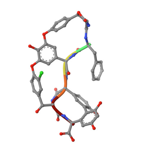
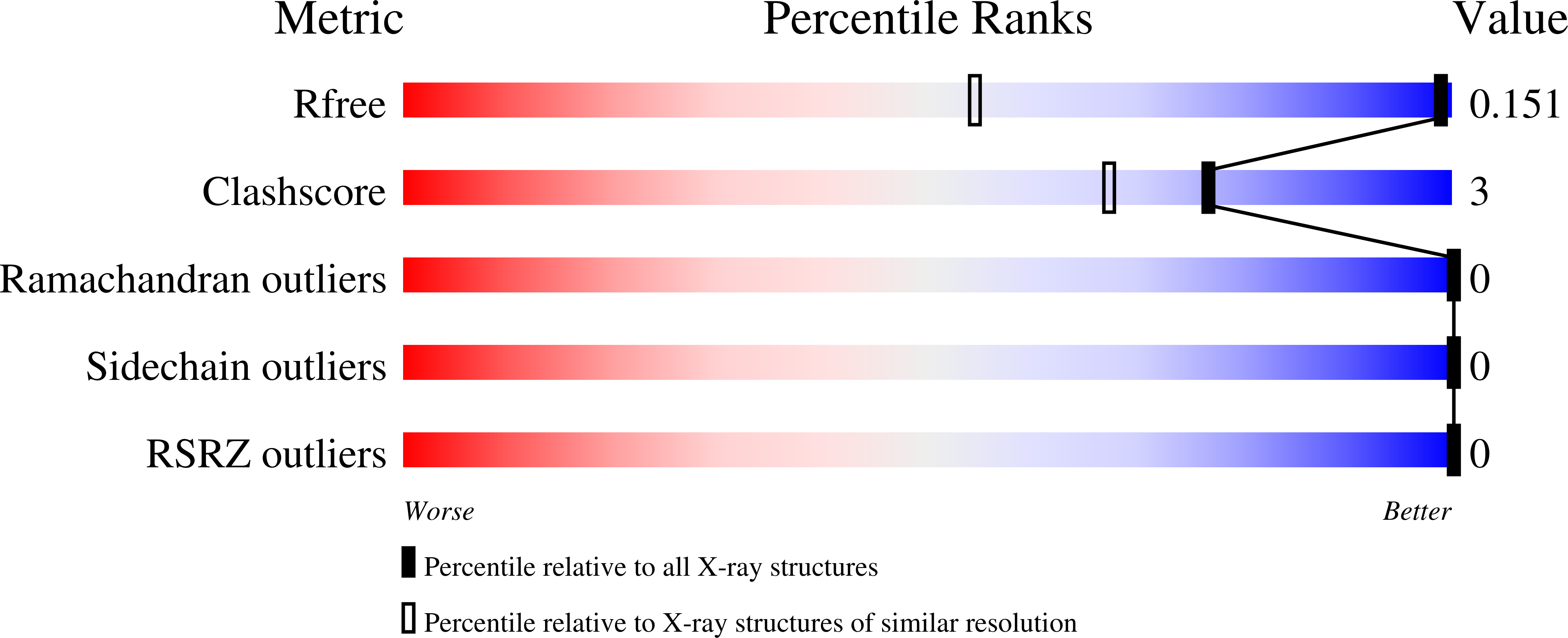
    7LKC, 7LTB

  • PubMed Abstract: 

    Keratinicyclins and keratinimicins are recently discovered glycopeptide antibiotics. Keratinimicins show broad-spectrum activity against Gram-positive bacteria, while keratinicyclins form a new chemotype by virtue of an unusual oxazolidinone moiety and exhibit specific antibiosis against Clostridioides difficile. Here we report the mechanism of action of keratinicyclin B (KCB). We find that steric constraints preclude KCB from binding peptidoglycan termini. Instead, KCB inhibits C. difficile growth by binding wall teichoic acids (WTAs) and interfering with cell wall remodeling. A computational model, guided by biochemical studies, provides an image of the interaction of KCB with C. difficile WTAs and shows that the same H-bonding framework used by glycopeptide antibiotics to bind peptidoglycan termini is used by KCB for interacting with WTAs. Analysis of KCB in combination with vancomycin (VAN) shows highly synergistic and specific antimicrobial activity, and that nanomolar combinations of the two drugs are sufficient for complete growth inhibition of C. difficile, while leaving common commensal strains unaffected.


  • Organizational Affiliation

    Department of Chemistry, Princeton University, Princeton, NJ, USA.


Macromolecules

Find similar proteins by:  Sequence   |   3D Structure  

Entity ID: 1
MoleculeChains Sequence LengthOrganismDetailsImage
Keratinicyclin B peptide moiety
A, B
6Amycolatopsis keratiniphilaMutation(s): 0 
Glycosylation
Glycosylation Sites: 1
Sequence Annotations
Expand
  • Reference Sequence
Oligosaccharides

Help

Entity ID: 2
MoleculeChains Length2D Diagram Glycosylation3D Interactions
3-ammonio-2,3,6-trideoxy-alpha-L-arabino-hexopyranose-(1-2)-beta-D-glucopyranoseC [auth F],
D [auth J]
2N/A
Experimental Data & Validation

Experimental Data

  • Method: X-RAY DIFFRACTION
  • Resolution: 0.95 Å
  • R-Value Free: 0.151 
  • R-Value Work: 0.146 
  • R-Value Observed: 0.146 
  • Space Group: P 21 21 21
Unit Cell:
Length ( Å )Angle ( ˚ )
a = 24.316α = 90
b = 29.667β = 90
c = 32.209γ = 90
Software Package:
Software NamePurpose
Aimlessdata scaling
PHASERphasing
PHENIXrefinement
PDB_EXTRACTdata extraction
XDSdata reduction

Structure Validation

View Full Validation Report


Ligand Structure Quality Assessment 


Entry History & Funding Information

Deposition Data


Funding OrganizationLocationGrant Number
National Institutes of Health/National Institute of General Medical Sciences (NIH/NIGMS)United StatesR01GM129496
National Institutes of Health/National Institute of General Medical Sciences (NIH/NIGMS)United States4R00GM129460

Revision History  (Full details and data files)

  • Version 1.0: 2022-12-07
    Type: Initial release
  • Version 1.1: 2023-10-25
    Changes: Data collection, Refinement description
  • Version 2.0: 2023-11-15
    Changes: Atomic model, Data collection, Derived calculations
  • Version 2.1: 2024-07-24
    Changes: Database references