7N2M

Crystal structure of DNA polymerase alpha catalytic core in complex with dCTP and template/primer having T-C mismatch at the post-insertion site


Experimental Data Snapshot

  • Method: X-RAY DIFFRACTION
  • Resolution: 2.90 Å
  • R-Value Free: 0.228 
  • R-Value Work: 0.195 

Starting Model: experimental
View more details

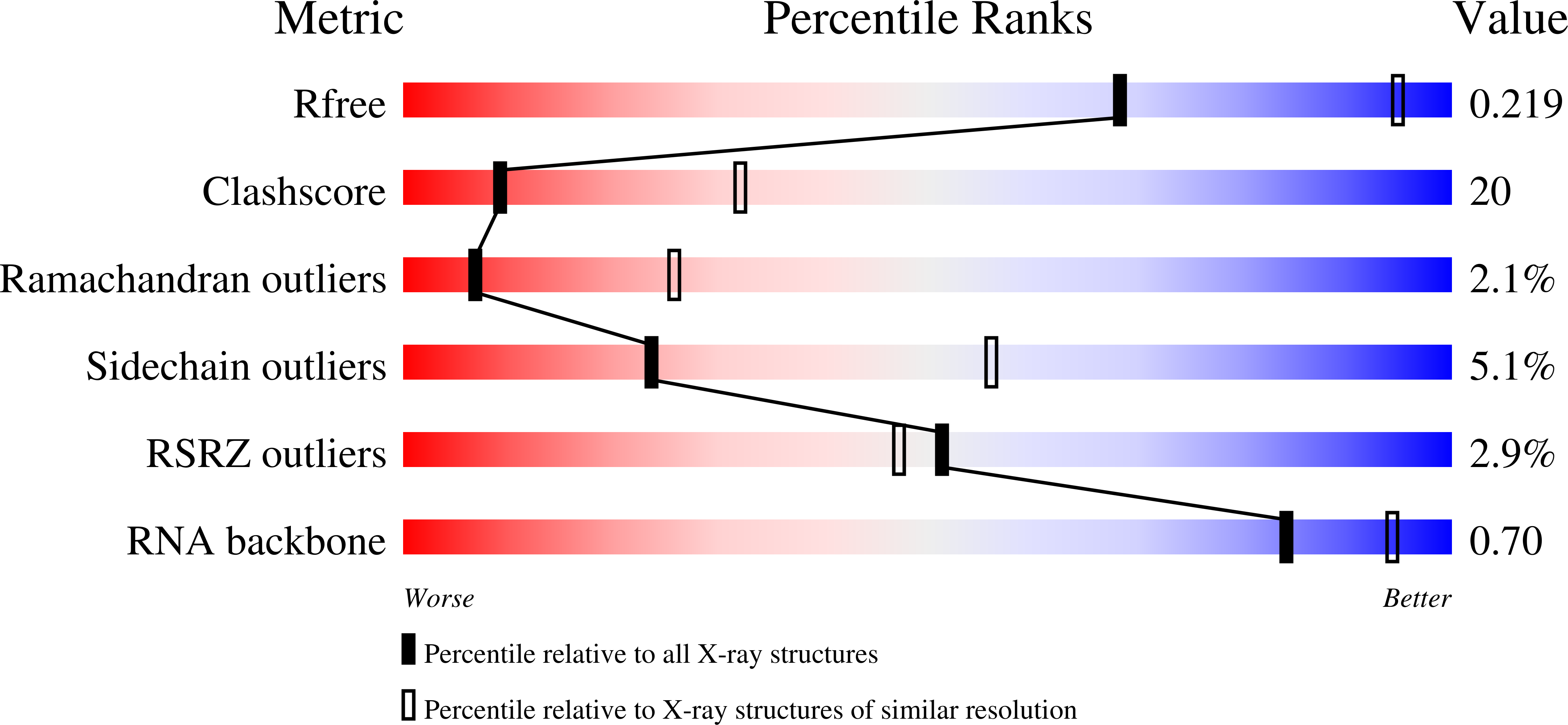
wwPDB Validation   3D Report Full Report


Ligand Structure Quality Assessment 


This is version 1.2 of the entry. See complete history


Literature

Structural and functional insight into mismatch extension by human DNA polymerase alpha.

Baranovskiy, A.G.Babayeva, N.D.Lisova, A.E.Morstadt, L.M.Tahirov, T.H.

(2022) Proc Natl Acad Sci U S A 119: e2111744119-e2111744119

  • DOI: https://doi.org/10.1073/pnas.2111744119
  • Primary Citation of Related Structures:  
    7N2M

  • PubMed Abstract: 

    Human DNA polymerase α (Polα) does not possess proofreading ability and plays an important role in genome replication and mutagenesis. Polα extends the RNA primers generated by primase and provides a springboard for loading other replication factors. Here we provide the structural and functional analysis of the human Polα interaction with a mismatched template:primer. The structure of the human Polα catalytic domain in the complex with an incoming deoxycytidine triphosphate (dCTP) and the template:primer containing a T-C mismatch at the growing primer terminus was solved at a 2.9 Å resolution. It revealed the absence of significant distortions in the active site and in the conformation of the substrates, except the primer 3′-end. The T-C mismatch acquired a planar geometry where both nucleotides moved toward each other by 0.4 Å and 0.7 Å, respectively, and made one hydrogen bond. The binding studies conducted at a physiological salt concentration revealed that Polα has a low affinity to DNA and is not able to discriminate against a mispaired template:primer in the absence of deoxynucleotide triphosphate (dNTP). Strikingly, in the presence of cognate dNTP, Polα showed a more than 10-fold higher selectivity for a correct duplex versus a mismatched one. According to pre-steady-state kinetic studies, human Polα extends the T-C mismatch with a 249-fold lower efficiency due to reduction of the polymerization rate constant by 38-fold and reduced affinity to the incoming nucleotide by 6.6-fold. Thus, a mismatch at the postinsertion site affects all factors important for primer extension: affinity to both substrates and the rate of DNA polymerization.


  • Organizational Affiliation

    Eppley Institute for Research in Cancer and Allied Diseases, Fred & Pamela Buffett Cancer Center, University of Nebraska Medical Center, Omaha, NE 68198.


Macromolecules

Find similar proteins by:  (by identity cutoff)  |  3D Structure
Entity ID: 1
MoleculeChains Sequence LengthOrganismDetailsImage
DNA polymerase alpha catalytic subunit922Homo sapiensMutation(s): 0 
Gene Names: POLA1POLA
EC: 2.7.7.7
UniProt & NIH Common Fund Data Resources
Find proteins for P09884 (Homo sapiens)
Explore P09884 
Go to UniProtKB:  P09884
PHAROS:  P09884
GTEx:  ENSG00000101868 
Entity Groups  
Sequence Clusters30% Identity50% Identity70% Identity90% Identity95% Identity100% Identity
UniProt GroupP09884
Sequence Annotations
Expand
  • Reference Sequence

Find similar nucleic acids by:  Sequence   |   3D Structure  

Entity ID: 2
MoleculeChains LengthOrganismImage
RNA (5'-R(*GP*CP*CP*UP*GP*GP*AP*GP*CP*GP*C)-3')11Homo sapiens
Sequence Annotations
Expand
  • Reference Sequence

Find similar nucleic acids by:  Sequence   |   3D Structure  

Entity ID: 3
MoleculeChains LengthOrganismImage
DNA (5'-D(*AP*T*AP*GP*TP*CP*GP*CP*TP*CP*CP*AP*GP*GP*C)-3')15Homo sapiens
Sequence Annotations
Expand
  • Reference Sequence
Small Molecules
Ligands 6 Unique
IDChains Name / Formula / InChI Key2D Diagram3D Interactions
DCP
Query on DCP

Download Ideal Coordinates CCD File 
D [auth A]2'-DEOXYCYTIDINE-5'-TRIPHOSPHATE
C9 H16 N3 O13 P3
RGWHQCVHVJXOKC-SHYZEUOFSA-N
1PE
Query on 1PE

Download Ideal Coordinates CCD File 
I [auth A]PENTAETHYLENE GLYCOL
C10 H22 O6
JLFNLZLINWHATN-UHFFFAOYSA-N
SO4
Query on SO4

Download Ideal Coordinates CCD File 
J [auth A]SULFATE ION
O4 S
QAOWNCQODCNURD-UHFFFAOYSA-L
ZN
Query on ZN

Download Ideal Coordinates CCD File 
F [auth A],
G [auth A],
K [auth B],
L [auth C]
ZINC ION
Zn
PTFCDOFLOPIGGS-UHFFFAOYSA-N
K
Query on K

Download Ideal Coordinates CCD File 
H [auth A]POTASSIUM ION
K
NPYPAHLBTDXSSS-UHFFFAOYSA-N
MG
Query on MG

Download Ideal Coordinates CCD File 
E [auth A]MAGNESIUM ION
Mg
JLVVSXFLKOJNIY-UHFFFAOYSA-N
Experimental Data & Validation

Experimental Data

  • Method: X-RAY DIFFRACTION
  • Resolution: 2.90 Å
  • R-Value Free: 0.228 
  • R-Value Work: 0.195 
  • Space Group: P 32 2 1
Unit Cell:
Length ( Å )Angle ( ˚ )
a = 140.285α = 90
b = 140.285β = 90
c = 181.066γ = 120
Software Package:
Software NamePurpose
HKL-2000data reduction
HKL-2000data scaling
CNSphasing
CNSrefinement
PDB_EXTRACTdata extraction

Structure Validation

View Full Validation Report


Ligand Structure Quality Assessment 


Entry History & Funding Information

Deposition Data


Funding OrganizationLocationGrant Number
National Institutes of Health/National Institute of General Medical Sciences (NIH/NIGMS)United StatesR35GM127085

Revision History  (Full details and data files)

  • Version 1.0: 2022-04-20
    Type: Initial release
  • Version 1.1: 2022-06-29
    Changes: Database references
  • Version 1.2: 2023-10-18
    Changes: Data collection, Refinement description