7NR4

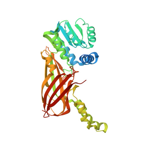
X-RAY STRUCTURE OF PRMT6 IN COMPLEX WITH indazole type inhibitor


Experimental Data Snapshot

  • Method: X-RAY DIFFRACTION
  • Resolution: 2.03 Å
  • R-Value Free: 0.235 
  • R-Value Work: 0.198 
  • R-Value Observed: 0.199 

Starting Model: experimental
View more details

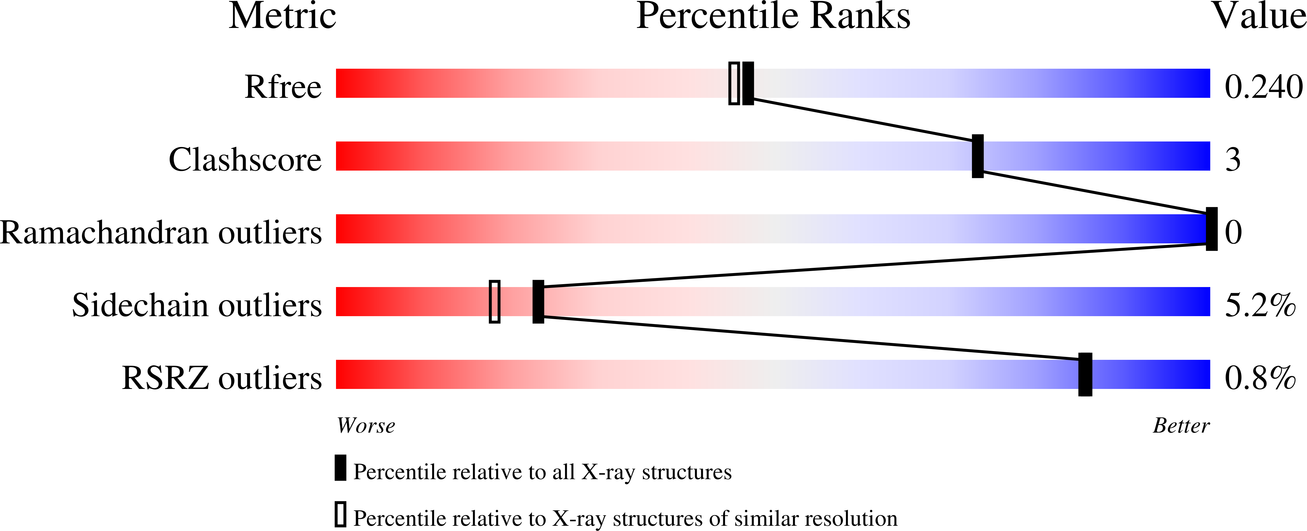
wwPDB Validation   3D Report Full Report


Ligand Structure Quality Assessment 


This is version 1.2 of the entry. See complete history


Literature

Rational Design and Synthesis of Selective PRMT4 Inhibitors: A New Chemotype for Development of Cancer Therapeutics*.

Sutherland, M.Li, A.Kaghad, A.Panagopoulos, D.Li, F.Szewczyk, M.Smil, D.Scholten, C.Bouche, L.Stellfeld, T.Arrowsmith, C.H.Barsyte, D.Vedadi, M.Hartung, I.V.Steuber, H.Britton, R.Santhakumar, V.

(2021) ChemMedChem 16: 1116-1125

  • DOI: https://doi.org/10.1002/cmdc.202100018
  • Primary Citation of Related Structures:  
    7NR4

  • PubMed Abstract: 

    Protein arginine N-methyl transferase 4 (PRMT4) asymmetrically dimethylates the arginine residues of histone H3 and nonhistone proteins. The overexpression of PRMT4 in several cancers has stimulated interest in the discovery of inhibitors as biological tools and, potentially, therapeutics. Although several PRMT4 inhibitors have been reported, most display poor selectivity against other members of the PRMT family of methyl transferases. Herein, we report the structure-based design of a new class of alanine-containing 3-arylindoles as potent and selective PRMT4 inhibitors, and describe key structure-activity relationships for this class of compounds.


  • Organizational Affiliation

    Department of Chemistry, Simon Fraser University, 8888 University Drive, Burnaby, BC V5A 1S6, Canada.


Macromolecules
Find similar proteins by:  (by identity cutoff)  |  3D Structure
Entity ID: 1
MoleculeChains Sequence LengthOrganismDetailsImage
Protein arginine N-methyltransferase 6
A, B, C, D
376Homo sapiensMutation(s): 0 
Gene Names: PRMT6HRMT1L6
EC: 2.1.1 (PDB Primary Data), 2.1.1.125 (PDB Primary Data), 2.1.1.319 (UniProt)
UniProt & NIH Common Fund Data Resources
Find proteins for Q96LA8 (Homo sapiens)
Explore Q96LA8 
Go to UniProtKB:  Q96LA8
PHAROS:  Q96LA8
GTEx:  ENSG00000198890 
Entity Groups  
Sequence Clusters30% Identity50% Identity70% Identity90% Identity95% Identity100% Identity
UniProt GroupQ96LA8
Sequence Annotations
Expand
  • Reference Sequence
Small Molecules
Ligands 2 Unique
IDChains Name / Formula / InChI Key2D Diagram3D Interactions
UO2 (Subject of Investigation/LOI)
Query on UO2

Download Ideal Coordinates CCD File 
F [auth A],
H [auth B],
J [auth C],
L [auth D]
(2~{S})-2-azanyl-~{N}-[3-[3-(dimethylsulfamoyl)phenyl]-2~{H}-indazol-5-yl]propanamide
C18 H21 N5 O3 S
HKVYLIVZRUYPKC-NSHDSACASA-N
SAH
Query on SAH

Download Ideal Coordinates CCD File 
E [auth A],
G [auth B],
I [auth C],
K [auth D]
S-ADENOSYL-L-HOMOCYSTEINE
C14 H20 N6 O5 S
ZJUKTBDSGOFHSH-WFMPWKQPSA-N
Experimental Data & Validation

Experimental Data

  • Method: X-RAY DIFFRACTION
  • Resolution: 2.03 Å
  • R-Value Free: 0.235 
  • R-Value Work: 0.198 
  • R-Value Observed: 0.199 
  • Space Group: P 1 21 1
Unit Cell:
Length ( Å )Angle ( ˚ )
a = 77.936α = 90
b = 135.252β = 98.28
c = 83.197γ = 90
Software Package:
Software NamePurpose
REFMACrefinement
PDB_EXTRACTdata extraction
XDSdata reduction
XDSdata scaling
PHASERphasing

Structure Validation

View Full Validation Report


Ligand Structure Quality Assessment 


Entry History 

Deposition Data

  • Released Date: 2021-04-07 
  • Deposition Author(s): Steuber, H.

Revision History  (Full details and data files)

  • Version 1.0: 2021-04-07
    Type: Initial release
  • Version 1.1: 2021-04-21
    Changes: Database references
  • Version 1.2: 2024-01-31
    Changes: Data collection, Database references, Refinement description