7NYT

Trichoderma reesei Cel7A E212Q mutant in complex with lactose.


Experimental Data Snapshot

  • Method: X-RAY DIFFRACTION
  • Resolution: 1.09 Å
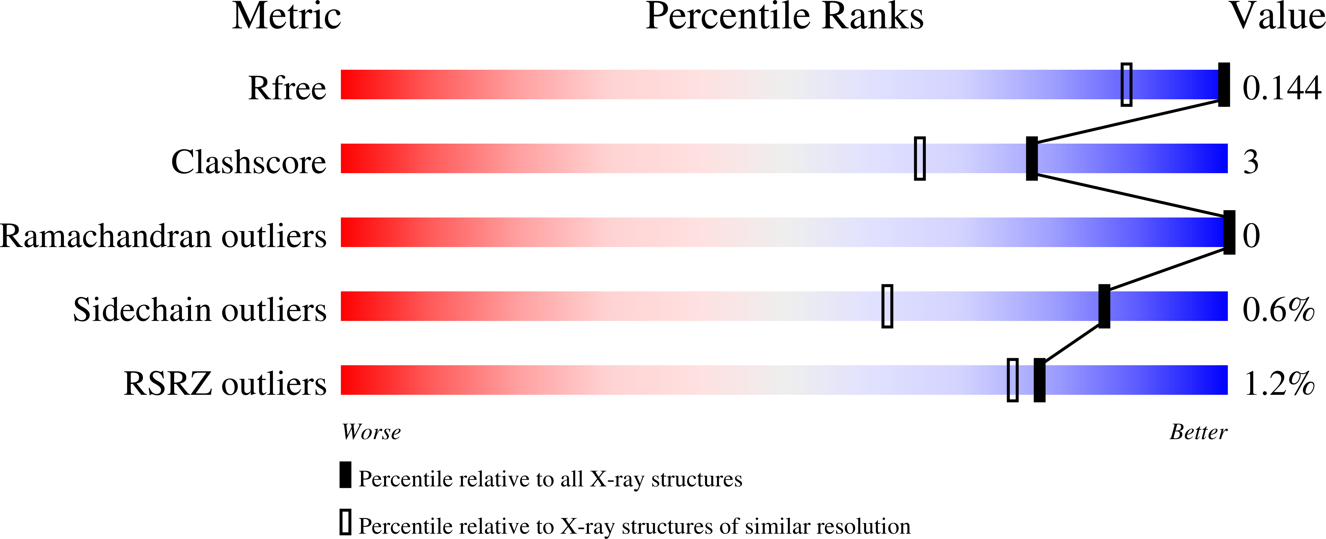
  • R-Value Free: 0.142 
  • R-Value Work: 0.123 
  • R-Value Observed: 0.124 

Starting Model: experimental
View more details

wwPDB Validation   3D Report Full Report


Ligand Structure Quality Assessment 


This is version 1.5 of the entry. See complete history


Literature

Enzyme kinetics by GH7 cellobiohydrolases on chromogenic substrates is dictated by non-productive binding: insights from crystal structures and MD simulation.

Haataja, T.Gado, J.E.Nutt, A.Anderson, N.T.Nilsson, M.Momeni, M.H.Isaksson, R.Valjamae, P.Johansson, G.Payne, C.M.Stahlberg, J.

(2023) FEBS J 290: 379-399

  • DOI: https://doi.org/10.1111/febs.16602
  • Primary Citation of Related Structures:  
    4UWT, 4V0Z, 7NYT, 7OC8

  • PubMed Abstract: 

    Cellobiohydrolases (CBHs) in the glycoside hydrolase family 7 (GH7) (EC3.2.1.176) are the major cellulose degrading enzymes both in industrial settings and in the context of carbon cycling in nature. Small carbohydrate conjugates such as p-nitrophenyl-β-d-cellobioside (pNPC), p-nitrophenyl-β-d-lactoside (pNPL) and methylumbelliferyl-β-d-cellobioside have commonly been used in colorimetric and fluorometric assays for analysing activity of these enzymes. Despite the similar nature of these compounds the kinetics of their enzymatic hydrolysis vary greatly between the different compounds as well as among different enzymes within the GH7 family. Through enzyme kinetics, crystallographic structure determination, molecular dynamics simulations, and fluorometric binding studies using the closely related compound o-nitrophenyl-β-d-cellobioside (oNPC), in this work we examine the different hydrolysis characteristics of these compounds on two model enzymes of this class, TrCel7A from Trichoderma reesei and PcCel7D from Phanerochaete chrysosporium. Protein crystal structures of the E212Q mutant of TrCel7A with pNPC and pNPL, and the wildtype TrCel7A with oNPC, reveal that non-productive binding at the product site is the dominating binding mode for these compounds. Enzyme kinetics results suggest the strength of non-productive binding is a key determinant for the activity characteristics on these substrates, with PcCel7D consistently showing higher turnover rates (k cat ) than TrCel7A, but higher Michaelis-Menten (K M ) constants as well. Furthermore, oNPC turned out to be useful as an active-site probe for fluorometric determination of the dissociation constant for cellobiose on TrCel7A but could not be utilized for the same purpose on PcCel7D, likely due to strong binding to an unknown site outside the active site.


  • Organizational Affiliation

    Department of Molecular Sciences, Swedish University of Agricultural Sciences, Uppsala, Sweden.


Macromolecules
Find similar proteins by:  (by identity cutoff)  |  3D Structure
Entity ID: 1
MoleculeChains Sequence LengthOrganismDetailsImage
Exoglucanase 1434Trichoderma reeseiMutation(s): 1 
Gene Names: cbh1
EC: 3.2.1.91
UniProt
Find proteins for P62694 (Hypocrea jecorina)
Explore P62694 
Go to UniProtKB:  P62694
Entity Groups  
Sequence Clusters30% Identity50% Identity70% Identity90% Identity95% Identity100% Identity
UniProt GroupP62694
Glycosylation
Glycosylation Sites: 1
Sequence Annotations
Expand
  • Reference Sequence
Oligosaccharides

Help

Entity ID: 2
MoleculeChains Length2D Diagram Glycosylation3D Interactions
beta-D-galactopyranose-(1-4)-beta-D-glucopyranose
B
2N/A
Glycosylation Resources
GlyTouCan:  G84224TW
GlyCosmos:  G84224TW
GlyGen:  G84224TW
Small Molecules
Ligands 5 Unique
IDChains Name / Formula / InChI Key2D Diagram3D Interactions
NAG
Query on NAG

Download Ideal Coordinates CCD File 
C [auth A]2-acetamido-2-deoxy-beta-D-glucopyranose
C8 H15 N O6
OVRNDRQMDRJTHS-FMDGEEDCSA-N
GAL (Subject of Investigation/LOI)
Query on GAL

Download Ideal Coordinates CCD File 
E [auth A]beta-D-galactopyranose
C6 H12 O6
WQZGKKKJIJFFOK-FPRJBGLDSA-N
BGC (Subject of Investigation/LOI)
Query on BGC

Download Ideal Coordinates CCD File 
D [auth A]beta-D-glucopyranose
C6 H12 O6
WQZGKKKJIJFFOK-VFUOTHLCSA-N
NPO (Subject of Investigation/LOI)
Query on NPO

Download Ideal Coordinates CCD File 
F [auth A]P-NITROPHENOL
C6 H5 N O3
BTJIUGUIPKRLHP-UHFFFAOYSA-N
CO
Query on CO

Download Ideal Coordinates CCD File 
G [auth A],
H [auth A]
COBALT (II) ION
Co
XLJKHNWPARRRJB-UHFFFAOYSA-N
Modified Residues  1 Unique
IDChains TypeFormula2D DiagramParent
PCA
Query on PCA
A
L-PEPTIDE LINKINGC5 H7 N O3GLN
Biologically Interesting Molecules (External Reference) 1 Unique
Experimental Data & Validation

Experimental Data

  • Method: X-RAY DIFFRACTION
  • Resolution: 1.09 Å
  • R-Value Free: 0.142 
  • R-Value Work: 0.123 
  • R-Value Observed: 0.124 
  • Space Group: I 2 2 2
Unit Cell:
Length ( Å )Angle ( ˚ )
a = 83.54α = 90
b = 82.21β = 90
c = 110.734γ = 90
Software Package:
Software NamePurpose
REFMACrefinement
REFMACphasing
pointlessdata processing
Aimlessdata scaling
pointlessdata processing
XDSdata reduction
EDNAdata processing
MxCuBEdata collection

Structure Validation

View Full Validation Report


Ligand Structure Quality Assessment 


Entry History & Funding Information

Deposition Data


Funding OrganizationLocationGrant Number
Swedish Energy AgencySweden42035-1

Revision History  (Full details and data files)

  • Version 1.0: 2022-03-09
    Type: Initial release
  • Version 1.1: 2022-09-21
    Changes: Database references
  • Version 1.2: 2022-12-07
    Changes: Structure summary
  • Version 1.3: 2023-02-01
    Changes: Database references
  • Version 1.4: 2024-02-07
    Changes: Data collection, Refinement description
  • Version 1.5: 2024-11-20
    Changes: Structure summary