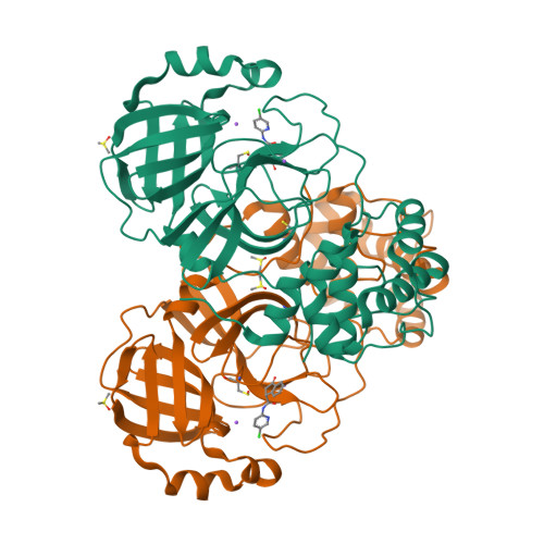
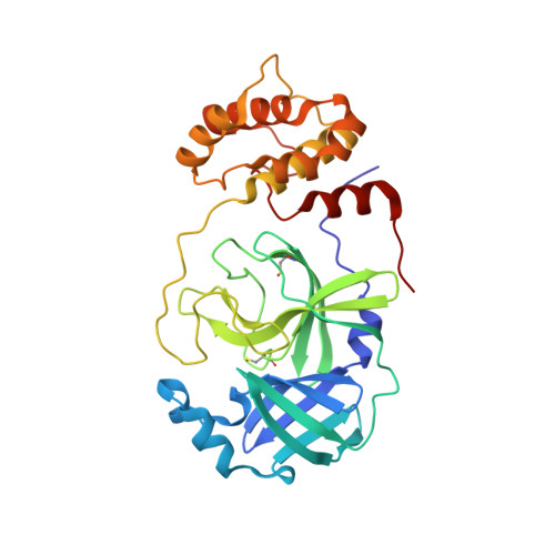
7P51

CRYSTAL STRUCTURE OF THE SARS-COV-2 MAIN PROTEASE COMPLEXED WITH FRAGMENT F01


Experimental Data Snapshot

  • Method: X-RAY DIFFRACTION
  • Resolution: 1.47 Å
  • R-Value Free: 0.216 
  • R-Value Work: 0.181 

Starting Model: experimental
View more details

wwPDB Validation   3D Report Full Report


Ligand Structure Quality Assessment 


This is version 1.3 of the entry. See complete history


Literature

NMR Spectroscopy of the Main Protease of SARS-CoV-2 and Fragment-Based Screening Identify Three Protein Hotspots and an Antiviral Fragment.

Cantrelle, F.X.Boll, E.Brier, L.Moschidi, D.Belouzard, S.Landry, V.Leroux, F.Dewitte, F.Landrieu, I.Dubuisson, J.Deprez, B.Charton, J.Hanoulle, X.

(2021) Angew Chem Int Ed Engl 60: 25428-25435

  • DOI: https://doi.org/10.1002/anie.202109965
  • Primary Citation of Related Structures:  
    7NTS, 7P51

  • PubMed Abstract: 

    The main protease (3CLp) of the SARS-CoV-2, the causative agent for the COVID-19 pandemic, is one of the main targets for drug development. To be active, 3CLp relies on a complex interplay between dimerization, active site flexibility, and allosteric regulation. The deciphering of these mechanisms is a crucial step to enable the search for inhibitors. In this context, using NMR spectroscopy, we studied the conformation of dimeric 3CLp from the SARS-CoV-2 and monitored ligand binding, based on NMR signal assignments. We performed a fragment-based screening that led to the identification of 38 fragment hits. Their binding sites showed three hotspots on 3CLp, two in the substrate binding pocket and one at the dimer interface. F01 is a non-covalent inhibitor of the 3CLp and has antiviral activity in SARS-CoV-2 infected cells. This study sheds light on the complex structure-function relationships of 3CLp and constitutes a strong basis to assist in developing potent 3CLp inhibitors.


  • Organizational Affiliation

    CNRS ERL9002-BSI-Integrative Structural Biology, 50 avenue Halley, F-59658 Villeneuve d'Ascq, Lille, France.


Macromolecules
Find similar proteins by:  (by identity cutoff)  |  3D Structure
Entity ID: 1
MoleculeChains Sequence LengthOrganismDetailsImage
3C-like proteinase306Severe acute respiratory syndrome coronavirus 2Mutation(s): 0 
EC: 3.4.22.69
UniProt
Find proteins for P0DTC1 (Severe acute respiratory syndrome coronavirus 2)
Explore P0DTC1 
Go to UniProtKB:  P0DTC1
Entity Groups  
Sequence Clusters30% Identity50% Identity70% Identity90% Identity95% Identity100% Identity
UniProt GroupP0DTC1
Sequence Annotations
Expand
  • Reference Sequence
Small Molecules
Modified Residues  1 Unique
IDChains TypeFormula2D DiagramParent
CSO
Query on CSO
A
L-PEPTIDE LINKINGC3 H7 N O3 SCYS
Experimental Data & Validation

Experimental Data

  • Method: X-RAY DIFFRACTION
  • Resolution: 1.47 Å
  • R-Value Free: 0.216 
  • R-Value Work: 0.181 
  • Space Group: C 1 2 1
Unit Cell:
Length ( Å )Angle ( ˚ )
a = 115.17α = 90
b = 53.56β = 101.24
c = 44.88γ = 90
Software Package:
Software NamePurpose
REFMACrefinement
XDSdata reduction
Aimlessdata scaling
MOLREPphasing
MxCuBEdata collection

Structure Validation

View Full Validation Report


Ligand Structure Quality Assessment 


Entry History & Funding Information

Deposition Data


Funding OrganizationLocationGrant Number
Other governmentFranceI-site ULNE - 3CLPRO-SCREEN-NMR

Revision History  (Full details and data files)

  • Version 1.0: 2021-10-06
    Type: Initial release
  • Version 1.1: 2021-11-24
    Changes: Database references
  • Version 1.2: 2024-01-31
    Changes: Data collection, Derived calculations, Refinement description
  • Version 1.3: 2024-11-20
    Changes: Structure summary