A Multifaceted Hit-Finding Approach Reveals Novel LC3 Family Ligands.
Steffek, M., Helgason, E., Popovych, N., Rouge, L., Bruning, J.M., Li, K.S., Burdick, D.J., Cai, J., Crawford, T., Xue, J., Decurtins, W., Fang, C., Grubers, F., Holliday, M.J., Langley, A., Petersen, A., Satz, A.L., Song, A., Stoffler, D., Strebel, Q., Tom, J.Y.K., Skelton, N., Staben, S.T., Wichert, M., Mulvihill, M.M., Dueber, E.C.(2023) Biochemistry 62: 633-644
- PubMed: 34985287
- DOI: https://doi.org/10.1021/acs.biochem.1c00682
- Primary Citation of Related Structures:
7R9W, 7R9Z, 7RA0 - PubMed Abstract:
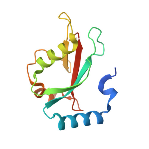
Autophagy-related proteins (Atgs) drive the lysosome-mediated degradation pathway, autophagy, to enable the clearance of dysfunctional cellular components and maintain homeostasis. In humans, this process is driven by the mammalian Atg8 (mAtg8) family of proteins comprising the LC3 and GABARAP subfamilies. The mAtg8 proteins play essential roles in the formation and maturation of autophagosomes and the capture of specific cargo through binding to the conserved LC3-interacting region (LIR) sequence within target proteins. Modulation of interactions of mAtg8 with its target proteins via small-molecule ligands would enable further interrogation of their function. Here we describe unbiased fragment and DNA-encoded library (DEL) screening approaches for discovering LC3 small-molecule ligands. Both strategies resulted in compounds that bind to LC3, with the fragment hits favoring a conserved hydrophobic pocket in mATG8 proteins, as detailed by LC3A-fragment complex crystal structures. Our findings demonstrate that the malleable LIR-binding surface can be readily targeted by fragments; however, rational design of additional interactions to drive increased affinity proved challenging. DEL libraries, which combine small, fragment-like building blocks into larger scaffolds, yielded higher-affinity binders and revealed an unexpected potential for reversible, covalent ligands. Moreover, DEL hits identified possible vectors for synthesizing fluorescent probes or bivalent molecules for engineering autophagic degradation of specific targets.
Organizational Affiliation:
Biochemical and Cellular Pharmacology, Genentech, 1 DNA Way, South San Francisco, California 94080, United States.


