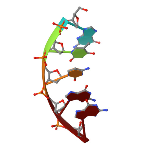
7RBG

Human DNA polymerase beta crosslinked ternary complex 1


Experimental Data Snapshot

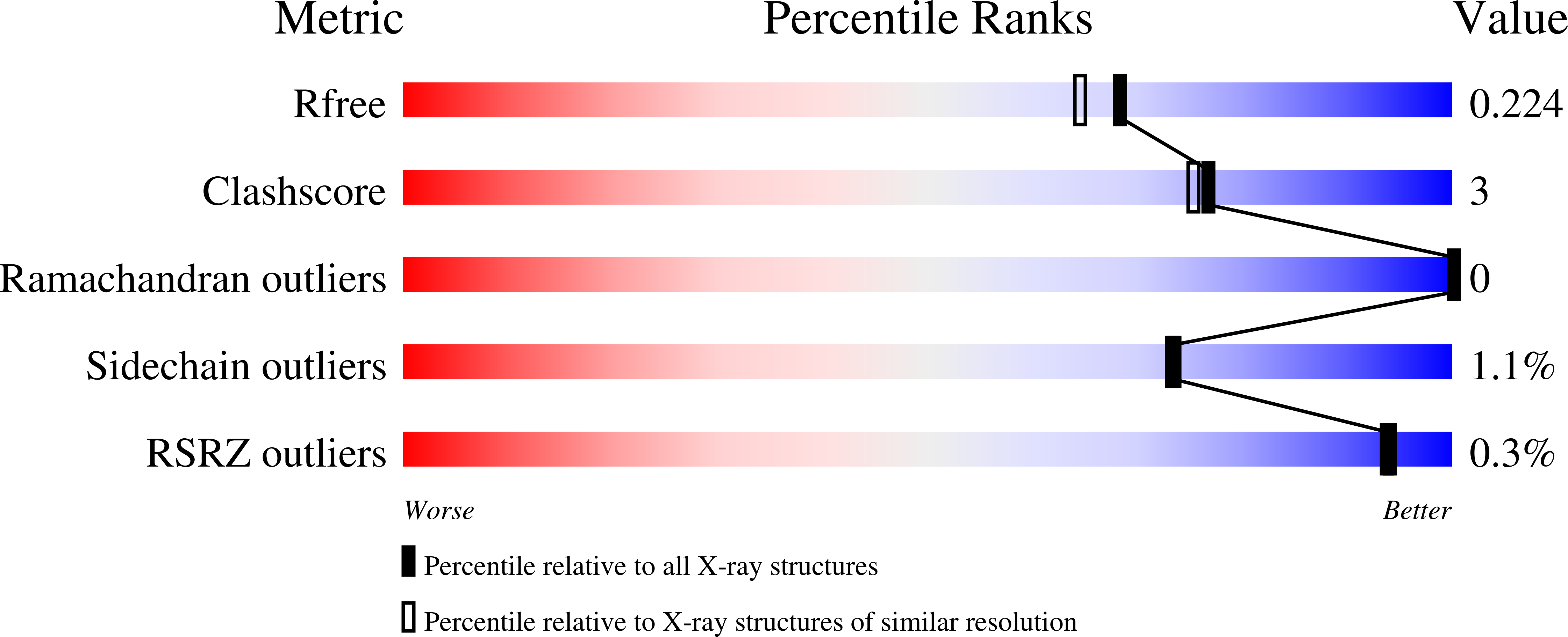
  • Method: X-RAY DIFFRACTION
  • Resolution: 1.90 Å
  • R-Value Free: 0.223 
  • R-Value Work: 0.179 
  • R-Value Observed: 0.181 

Starting Model: experimental
View more details

wwPDB Validation   3D Report Full Report


Ligand Structure Quality Assessment 


This is version 1.4 of the entry. See complete history


Literature

Interlocking activities of DNA polymerase beta in the base excision repair pathway.

Kumar, A.Reed, A.J.Zahurancik, W.J.Daskalova, S.M.Hecht, S.M.Suo, Z.

(2022) Proc Natl Acad Sci U S A 119: e2118940119-e2118940119

  • DOI: https://doi.org/10.1073/pnas.2118940119
  • Primary Citation of Related Structures:  
    7RBE, 7RBF, 7RBG, 7RBH, 7RBI, 7RBJ, 7RBK, 7RBL, 7RBM, 7RBN, 7RBO

  • PubMed Abstract: 

    SignificanceBase excision repair (BER) is one of the major DNA repair pathways used to fix a myriad of cellular DNA lesions. The enzymes involved in BER, including DNA polymerase β (Polβ), have been identified and characterized, but how they act together to efficiently perform BER has not been fully understood. Through gel electrophoresis, mass spectrometry, and kinetic analysis, we discovered that the two enzymatic activities of Polβ can be interlocked, rather than functioning independently from each other, when processing DNA intermediates formed in BER. The finding prompted us to hypothesize a modified BER pathway. Through conventional and time-resolved X-ray crystallography, we solved 11 high-resolution crystal structures of cross-linked Polβ complexes and proposed a detailed chemical mechanism for Polβ's 5'-deoxyribose-5-phosphate lyase activity.


  • Organizational Affiliation

    Department of Biomedical Sciences, Florida State University College of Medicine, Tallahassee, FL 32306.


Macromolecules

Find similar proteins by:  (by identity cutoff)  |  3D Structure
Entity ID: 4
MoleculeChains Sequence LengthOrganismDetailsImage
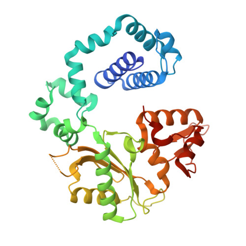
DNA polymerase betaD [auth A]341Homo sapiensMutation(s): 0 
Gene Names: POLB
EC: 2.7.7.7 (PDB Primary Data), 4.2.99 (PDB Primary Data), 4.2.99.18 (UniProt)
UniProt & NIH Common Fund Data Resources
Find proteins for P06746 (Homo sapiens)
Explore P06746 
Go to UniProtKB:  P06746
PHAROS:  P06746
GTEx:  ENSG00000070501 
Entity Groups  
Sequence Clusters30% Identity50% Identity70% Identity90% Identity95% Identity100% Identity
UniProt GroupP06746
Sequence Annotations
Expand
  • Reference Sequence

Find similar nucleic acids by:  Sequence   |   3D Structure  

Entity ID: 1
MoleculeChains LengthOrganismImage
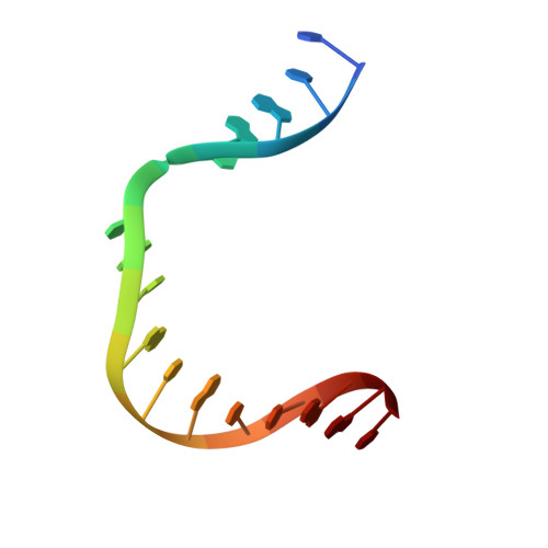
DNA (5'-D(*CP*CP*GP*AP*CP*GP*GP*CP*GP*CP*AP*TP*CP*AP*GP*C)-3')A [auth T]16synthetic construct
Sequence Annotations
Expand
  • Reference Sequence

Find similar nucleic acids by:  Sequence   |   3D Structure  

Entity ID: 2
MoleculeChains LengthOrganismImage
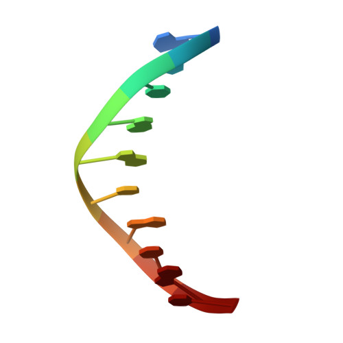
DNA (5'-D(*GP*CP*TP*GP*AP*TP*GP*CP*GP*C)-3')B [auth P]10synthetic construct
Sequence Annotations
Expand
  • Reference Sequence

Find similar nucleic acids by:  Sequence   |   3D Structure  

Entity ID: 3
MoleculeChains LengthOrganismImage
DNA (5'-D(*GP*TP*CP*GP*G)-3')C [auth D]5synthetic construct
Sequence Annotations
Expand
  • Reference Sequence
Small Molecules
Ligands 5 Unique
IDChains Name / Formula / InChI Key2D Diagram3D Interactions
DCP
Query on DCP

Download Ideal Coordinates CCD File 
E [auth A]2'-DEOXYCYTIDINE-5'-TRIPHOSPHATE
C9 H16 N3 O13 P3
RGWHQCVHVJXOKC-SHYZEUOFSA-N
QPJ (Subject of Investigation/LOI)
Query on QPJ

Download Ideal Coordinates CCD File 
F [auth A]2-deoxy-3,5-di-O-phosphono-D-erythro-pentitol
C5 H14 O10 P2
BUWHXMOYXFBKFR-UHNVWZDZSA-N
CA
Query on CA

Download Ideal Coordinates CCD File 
G [auth A],
H [auth A],
I [auth A]
CALCIUM ION
Ca
BHPQYMZQTOCNFJ-UHFFFAOYSA-N
CL
Query on CL

Download Ideal Coordinates CCD File 
L [auth A],
M [auth A]
CHLORIDE ION
Cl
VEXZGXHMUGYJMC-UHFFFAOYSA-M
NA
Query on NA

Download Ideal Coordinates CCD File 
J [auth A],
K [auth A]
SODIUM ION
Na
FKNQFGJONOIPTF-UHFFFAOYSA-N
Experimental Data & Validation

Experimental Data

  • Method: X-RAY DIFFRACTION
  • Resolution: 1.90 Å
  • R-Value Free: 0.223 
  • R-Value Work: 0.179 
  • R-Value Observed: 0.181 
  • Space Group: P 1 21 1
Unit Cell:
Length ( Å )Angle ( ˚ )
a = 55.51α = 90
b = 78.43β = 113.22
c = 57.19γ = 90
Software Package:
Software NamePurpose
Aimlessdata scaling
REFMACrefinement
PDB_EXTRACTdata extraction
XDSdata reduction
PHASERphasing

Structure Validation

View Full Validation Report


Ligand Structure Quality Assessment 


Entry History & Funding Information

Deposition Data


Funding OrganizationLocationGrant Number
National Institutes of Health/National Institute of General Medical Sciences (NIH/NIGMS)United StatesGM122093

Revision History  (Full details and data files)

  • Version 1.0: 2022-03-09
    Type: Initial release
  • Version 1.1: 2022-03-16
    Changes: Database references
  • Version 1.2: 2022-04-20
    Changes: Source and taxonomy
  • Version 1.3: 2023-10-18
    Changes: Data collection, Refinement description
  • Version 1.4: 2024-11-20
    Changes: Structure summary