7SKU | pdb_00007sku

Nipah virus matrix protein in complex with PI(4,5)P2


Experimental Data Snapshot

  • Method: X-RAY DIFFRACTION
  • Resolution: 2.12 Å
  • R-Value Free: 
    0.264 (Depositor), 0.250 (DCC) 
  • R-Value Work: 
    0.231 (Depositor), 0.230 (DCC) 
  • R-Value Observed: 
    0.233 (Depositor) 

Starting Model: experimental
View more details

wwPDB Validation   3D Report Full Report


Ligand Structure Quality Assessment 

Created with Raphaël 2.3.0Worse 01 BetterLigand structure goodness of fit to experimental dataBest fitted PIOClick on this verticalbar to view details

This is version 1.1 of the entry. See complete history


Literature

Measles and Nipah virus assembly: Specific lipid binding drives matrix polymerization.

Norris, M.J.Husby, M.L.Kiosses, W.B.Yin, J.Saxena, R.Rennick, L.J.Heiner, A.Harkins, S.S.Pokhrel, R.Schendel, S.L.Hastie, K.M.Landeras-Bueno, S.Salie, Z.L.Lee, B.Chapagain, P.P.Maisner, A.Duprex, W.P.Stahelin, R.V.Saphire, E.O.

(2022) Sci Adv 8: eabn1440-eabn1440

  • DOI: https://doi.org/10.1126/sciadv.abn1440
  • Primary Citation of Related Structures:  
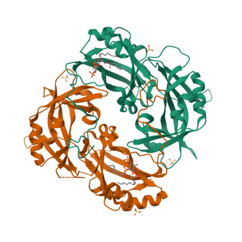
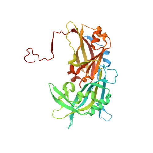
    7SKS, 7SKT, 7SKU

  • PubMed Abstract: 

    Measles virus, Nipah virus, and multiple other paramyxoviruses cause disease outbreaks in humans and animals worldwide. The paramyxovirus matrix (M) protein mediates virion assembly and budding from host cell membranes. M is thus a key target for antivirals, but few high-resolution structures of paramyxovirus M are available, and we lack the clear understanding of how viral M proteins interact with membrane lipids to mediate viral assembly and egress that is needed to guide antiviral design. Here, we reveal that M proteins associate with phosphatidylserine and phosphatidylinositol 4,5-bisphosphate [PI(4,5)P 2 ] at the plasma membrane. Using x-ray crystallography, electron microscopy, and molecular dynamics, we demonstrate that PI(4,5)P 2 binding induces conformational and electrostatic changes in the M protein surface that trigger membrane deformation, matrix layer polymerization, and virion assembly.


  • Organizational Affiliation

    Center for Infectious Disease and Vaccine Research, La Jolla Institute for Immunology, La Jolla, CA 92037, USA.


Macromolecules
Find similar proteins by:  (by identity cutoff)  |  3D Structure
Entity ID: 1
MoleculeChains Sequence LengthOrganismDetailsImage
Matrix protein
A, B
393Henipavirus nipahenseMutation(s): 0 
UniProt
Find proteins for Q9IK90 (Nipah virus)
Explore Q9IK90 
Go to UniProtKB:  Q9IK90
Entity Groups  
Sequence Clusters30% Identity50% Identity70% Identity90% Identity95% Identity100% Identity
UniProt GroupQ9IK90
Sequence Annotations
Expand
  • Reference Sequence
Experimental Data & Validation

Experimental Data

  • Method: X-RAY DIFFRACTION
  • Resolution: 2.12 Å
  • R-Value Free:  0.264 (Depositor), 0.250 (DCC) 
  • R-Value Work:  0.231 (Depositor), 0.230 (DCC) 
  • R-Value Observed: 0.233 (Depositor) 
Space Group: P 21 21 21
Unit Cell:
Length ( Å )Angle ( ˚ )
a = 70.114α = 90
b = 96.471β = 90
c = 119.972γ = 90
Software Package:
Software NamePurpose
autoPROCdata processing
XDSdata reduction
Aimlessdata scaling
TRUNCATEdata processing
BUSTERrefinement
PHASERphasing

Structure Validation

View Full Validation Report


Ligand Structure Quality Assessment 

Created with Raphaël 2.3.0Worse 01 BetterLigand structure goodness of fit to experimental dataBest fitted PIOClick on this verticalbar to view details

Entry History & Funding Information

Deposition Data


Funding OrganizationLocationGrant Number
Not fundedUnited States--

Revision History  (Full details and data files)

  • Version 1.0: 2022-08-24
    Type: Initial release
  • Version 1.1: 2023-10-18
    Changes: Data collection, Refinement description