Phenol-soluble modulins PSM alpha 3 and PSM beta 2 form nanotubes that are cross-alpha amyloids.
Kreutzberger, M.A.B., Wang, S., Beltran, L.C., Tuachi, A., Zuo, X., Egelman, E.H., Conticello, V.P.(2022) Proc Natl Acad Sci U S A 119: e2121586119-e2121586119
- PubMed: 35533283
- DOI: https://doi.org/10.1073/pnas.2121586119
- Primary Citation of Related Structures:
7SZZ, 7T0X, 7T8U - PubMed Abstract:
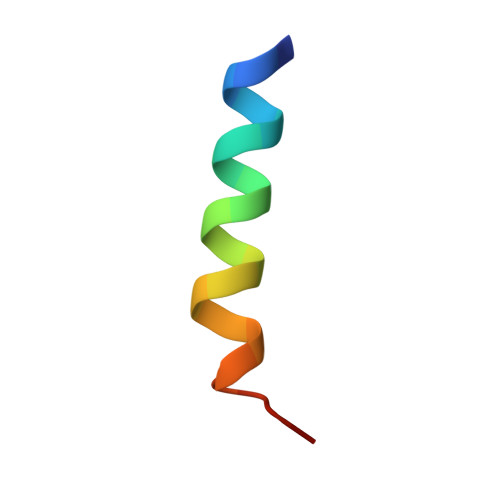
Phenol-soluble modulins (PSMs) are peptide-based virulence factors that play significant roles in the pathogenesis of staphylococcal strains in community-associated and hospital-associated infections. In addition to cytotoxicity, PSMs display the propensity to self-assemble into fibrillar species, which may be mediated through the formation of amphipathic conformations. Here, we analyze the self-assembly behavior of two PSMs, PSMα3 and PSMβ2, which are derived from peptides expressed by methicillin-resistant Staphylococcus aureus (MRSA), a significant human pathogen. In both cases, we observed the formation of a mixture of self-assembled species including twisted filaments, helical ribbons, and nanotubes, which can reversibly interconvert in vitro. Cryo–electron microscopy structural analysis of three PSM nanotubes, two derived from PSMα3 and one from PSMβ2, revealed that the assemblies displayed remarkably similar structures based on lateral association of cross-α amyloid protofilaments. The amphipathic helical conformations of PSMα3 and PSMβ2 enforced a bilayer arrangement within the protofilaments that defined the structures of the respective PSMα3 and PSMβ2 nanotubes. We demonstrate that, similar to amyloids based on cross-β protofilaments, cross-α amyloids derived from these PSMs display polymorphism, not only in terms of the global morphology (e.g., twisted filament, helical ribbon, and nanotube) but also with respect to the number of protofilaments within a given peptide assembly. These results suggest that the folding landscape of PSM derivatives may be more complex than originally anticipated and that the assemblies are able to sample a wide range of supramolecular structural space.
Organizational Affiliation:
Department of Biochemistry and Molecular Genetics, University of Virginia, Charlottesville, VA 22908.