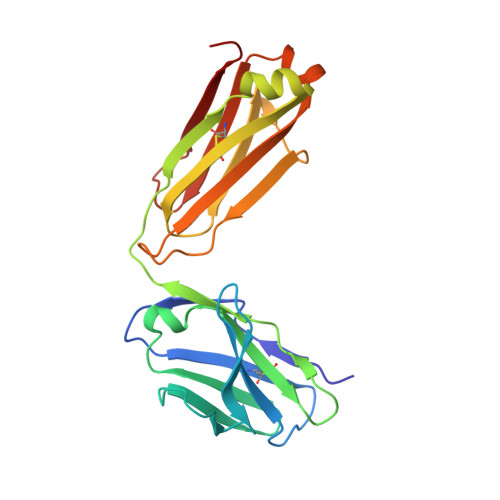
7UPW

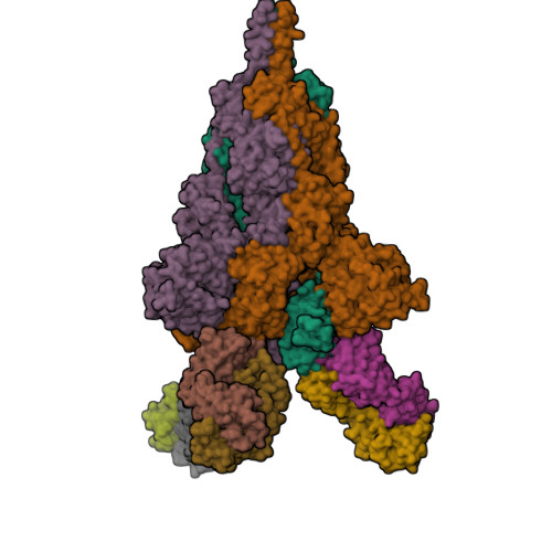
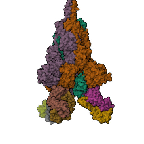
Three RBD-down state of SARS-CoV-2 D614G spike in complex with the SP1-77 neutralizing antibody Fab fragment


Experimental Data Snapshot

  • Method: ELECTRON MICROSCOPY
  • Resolution: 2.70 Å
  • Aggregation State: PARTICLE 
  • Reconstruction Method: SINGLE PARTICLE 

Starting Model: experimental
View more details

wwPDB Validation   3D Report Full Report


This is version 1.3 of the entry. See complete history


Literature

An antibody from single human V H -rearranging mouse neutralizes all SARS-CoV-2 variants through BA.5 by inhibiting membrane fusion.

Luo, S.Zhang, J.Kreutzberger, A.J.B.Eaton, A.Edwards, R.J.Jing, C.Dai, H.Q.Sempowski, G.D.Cronin, K.Parks, R.Ye, A.Y.Mansouri, K.Barr, M.Pishesha, N.Williams, A.C.Vieira Francisco, L.Saminathan, A.Peng, H.Batra, H.Bellusci, L.Khurana, S.Alam, S.M.Montefiori, D.C.Saunders, K.O.Tian, M.Ploegh, H.Kirchhausen, T.Chen, B.Haynes, B.F.Alt, F.W.

(2022) Sci Immunol 7: eadd5446-eadd5446

  • DOI: https://doi.org/10.1126/sciimmunol.add5446
  • Primary Citation of Related Structures:  
    7UPW, 7UPX, 7UPY

  • PubMed Abstract: 

    SARS-CoV-2 Omicron subvariants have generated a worldwide health crisis due to resistance to most approved SARS-CoV-2 neutralizing antibodies and evasion of vaccination-induced antibodies. To manage Omicron subvariants and prepare for new ones, additional means of isolating broad and potent humanized SARS-CoV-2 neutralizing antibodies are desirable. Here, we describe a mouse model in which the primary B cell receptor (BCR) repertoire is generated solely through V(D)J recombination of a human V H 1-2 heavy chain (HC) and, substantially, a human Vκ1-33 light chain (LC). Thus, primary humanized BCR repertoire diversity in these mice derives from immensely diverse HC and LC antigen-contact CDR3 sequences generated by nontemplated junctional modifications during V(D)J recombination. Immunizing this mouse model with SARS-CoV-2 (Wuhan-Hu-1) spike protein immunogens elicited several V H 1-2/Vκ1-33-based neutralizing antibodies that bound RBD in a different mode from each other and from those of many prior patient-derived V H 1-2-based neutralizing antibodies. Of these, SP1-77 potently and broadly neutralized all SARS-CoV-2 variants through BA.5. Cryo-EM studies revealed that SP1-77 bound RBD away from the receptor-binding motif via a CDR3-dominated recognition mode. Lattice light-sheet microscopy-based studies showed that SP1-77 did not block ACE2-mediated viral attachment or endocytosis but rather blocked viral-host membrane fusion. The broad and potent SP1-77 neutralization activity and nontraditional mechanism of action suggest that it might have therapeutic potential. Likewise, the SP1-77 binding epitope may inform vaccine strategies. Last, the type of humanized mouse models that we have described may contribute to identifying therapeutic antibodies against future SARS-CoV-2 variants and other pathogens.


  • Organizational Affiliation

    Howard Hughes Medical Institute, Program in Cellular and Molecular Medicine, Boston Children's Hospital, Boston, MA 02115, USA.


Macromolecules
Find similar proteins by:  (by identity cutoff)  |  3D Structure
Entity ID: 1
MoleculeChains Sequence LengthOrganismDetailsImage
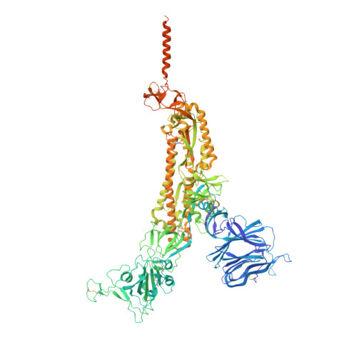
Spike glycoprotein
A, B, C
1,310Severe acute respiratory syndrome coronavirusMutation(s): 1 
Gene Names: S2
UniProt
Find proteins for P0DTC2 (Severe acute respiratory syndrome coronavirus 2)
Explore P0DTC2 
Go to UniProtKB:  P0DTC2
Entity Groups  
Sequence Clusters30% Identity50% Identity70% Identity90% Identity95% Identity100% Identity
UniProt GroupP0DTC2
Glycosylation
Glycosylation Sites: 19Go to GlyGen: P0DTC2-1
Sequence Annotations
Expand
  • Reference Sequence
Find similar proteins by:  (by identity cutoff)  |  3D Structure
Entity ID: 2
MoleculeChains Sequence LengthOrganismDetailsImage
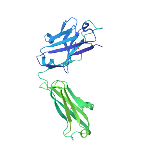
SP1-77 Fab heavy chain
D, F, H
451Homo sapiensMus musculusMutation(s): 0 
Entity Groups  
Sequence Clusters30% Identity50% Identity70% Identity90% Identity95% Identity100% Identity
Sequence Annotations
Expand
  • Reference Sequence
Find similar proteins by:  (by identity cutoff)  |  3D Structure
Entity ID: 3
MoleculeChains Sequence LengthOrganismDetailsImage
SP1-77 Fab light chainE,
G,
I [auth L]
213Homo sapiensMus musculusMutation(s): 0 
Entity Groups  
Sequence Clusters30% Identity50% Identity70% Identity90% Identity95% Identity100% Identity
Sequence Annotations
Expand
  • Reference Sequence
Oligosaccharides

Help

Entity ID: 4
MoleculeChains Length2D Diagram Glycosylation3D Interactions
2-acetamido-2-deoxy-beta-D-glucopyranose-(1-4)-2-acetamido-2-deoxy-beta-D-glucopyranose
HA [auth h],
J [auth I],
LA [auth l],
N,
V,
HA [auth h],
J [auth I],
LA [auth l],
N,
V,
Z
2N-Glycosylation
Glycosylation Resources
GlyTouCan:  G42666HT
GlyCosmos:  G42666HT
GlyGen:  G42666HT
Entity ID: 5
MoleculeChains Length2D Diagram Glycosylation3D Interactions
alpha-D-mannopyranose-(1-4)-2-acetamido-2-deoxy-beta-D-glucopyranose-(1-4)-2-acetamido-2-deoxy-beta-D-glucopyranose
AA [auth a],
BA [auth b],
DA [auth d],
EA [auth e],
GA [auth g],
AA [auth a],
BA [auth b],
DA [auth d],
EA [auth e],
GA [auth g],
IA [auth i],
JA [auth j],
K [auth J],
KA [auth k],
L [auth K],
M,
MA [auth m],
NA [auth n],
O,
P,
PA [auth p],
QA [auth q],
R,
S,
SA [auth s],
U,
W,
X,
Y
3N-Glycosylation
Glycosylation Resources
GlyTouCan:  G62182OO
GlyCosmos:  G62182OO
GlyGen:  G62182OO
Entity ID: 6
MoleculeChains Length2D Diagram Glycosylation3D Interactions
2-acetamido-2-deoxy-beta-D-glucopyranose-(1-4)-[alpha-L-fucopyranose-(1-6)]2-acetamido-2-deoxy-beta-D-glucopyranose
CA [auth c],
FA [auth f],
OA [auth o],
Q,
RA [auth r],
CA [auth c],
FA [auth f],
OA [auth o],
Q,
RA [auth r],
T
3N-Glycosylation
Glycosylation Resources
GlyTouCan:  G21290RB
GlyCosmos:  G21290RB
GlyGen:  G21290RB
Small Molecules
Ligands 1 Unique
IDChains Name / Formula / InChI Key2D Diagram3D Interactions
NAG (Subject of Investigation/LOI)
Query on NAG

Download Ideal Coordinates CCD File 
AB [auth B]
BB [auth B]
CB [auth B]
DB [auth B]
EB [auth B]
AB [auth B],
BB [auth B],
CB [auth B],
DB [auth B],
EB [auth B],
FB [auth B],
GB [auth B],
HB [auth C],
IB [auth C],
JB [auth C],
KB [auth C],
LB [auth C],
MB [auth C],
NB [auth C],
TA [auth A],
UA [auth A],
VA [auth A],
WA [auth A],
XA [auth A],
YA [auth A],
ZA [auth A]
2-acetamido-2-deoxy-beta-D-glucopyranose
C8 H15 N O6
OVRNDRQMDRJTHS-FMDGEEDCSA-N
Experimental Data & Validation

Experimental Data

  • Method: ELECTRON MICROSCOPY
  • Resolution: 2.70 Å
  • Aggregation State: PARTICLE 
  • Reconstruction Method: SINGLE PARTICLE 
EM Software:
TaskSoftware PackageVersion
MODEL REFINEMENTPHENIX
RECONSTRUCTIONcryoSPARC

Structure Validation

View Full Validation Report


Entry History & Funding Information

Deposition Data


Funding OrganizationLocationGrant Number
Howard Hughes Medical Institute (HHMI)United StatesINV-021989
National Institutes of Health/National Institute Of Allergy and Infectious Diseases (NIH/NIAID)United StatesAI27193
Massachusetts Consortium on Pathogen Readiness (MassCPR)United StatesSARS-CoV-2 Variants Award
Mercatus CenterUnited StatesEmergent Ventures Fast Grant

Revision History  (Full details and data files)

  • Version 1.0: 2022-08-10
    Type: Initial release
  • Version 1.1: 2022-12-14
    Changes: Refinement description
  • Version 1.2: 2023-08-23
    Changes: Data collection, Database references
  • Version 1.3: 2024-10-30
    Changes: Data collection, Refinement description, Structure summary