Experimental and theoretical study on converting myoglobin into a stable domain-swapped dimer by utilizing a tight hydrogen bond network at the hinge region.
Xie, C., Shimoyama, H., Yamanaka, M., Nagao, S., Komori, H., Shibata, N., Higuchi, Y., Shigeta, Y., Hirota, S.(2021) RSC Adv 11: 37604-37611
- PubMed: 35496441
- DOI: https://doi.org/10.1039/d1ra06888a
- Primary Citation of Related Structures:
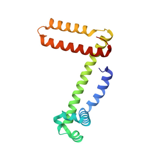
7V5P, 7V5Q, 7V5R - PubMed Abstract:
Various factors, such as helical propensity and hydrogen bonds, control protein structures. A frequently used model protein, myoglobin (Mb), can perform 3D domain swapping, in which the loop at the hinge region is converted to a helical structure in the dimer. We have previously succeeded in obtaining monomer-dimer equilibrium in the native state by introducing a high α-helical propensity residue, Ala, to the hinge region. In this study, we focused on another factor that governs the protein structure, hydrogen bonding. X-ray crystal structures and thermodynamic studies showed that the myoglobin dimer was stabilized over the monomer when keeping His82 to interact with Lys79 and Asp141 through water moleclues and mutating Leu137, which was located close to the H-bond network at the dimer hinge region, to a hydrophilic amino acid (Glu or Asp). Molecular dynamics simulation studies confirmed that the number of H-bonds increased and the α-helices at the hinge region became more rigid for mutants with a tighter H-bond network, supporting the hypothesis that the myoglobin dimer is stabilized when the H-bond network at the hinge region is enhanced. This demonstrates the importance and utility of hydrogen bonds for designing a protein dimer from its monomer with 3D domain swapping.
Organizational Affiliation:
Division of Materials Science, Graduate School of Science and Technology, Nara Institute of Science and Technology 8916-5 Takayama, Ikoma Nara 630-0192 Japan hirota@ms.naist.jp.