7XBG

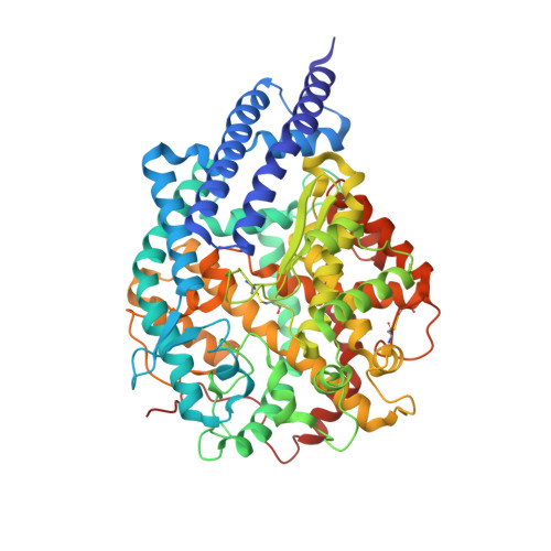
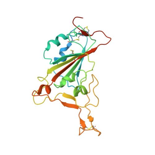
The crystal structure of RshSTT182/200 RBD-insert2-T346R-Y496G mutant in complex with human ACE2


Experimental Data Snapshot

  • Method: X-RAY DIFFRACTION
  • Resolution: 3.37 Å
  • R-Value Free: 0.274 
  • R-Value Work: 0.269 
  • R-Value Observed: 0.270 

Starting Model: experimental
View more details

wwPDB Validation   3D Report Full Report


Ligand Structure Quality Assessment 


This is version 1.3 of the entry. See complete history


Literature

Host range and structural analysis of bat-origin RshSTT182/200 coronavirus binding to human ACE2 and its animal orthologs.

Hu, Y.Liu, K.Han, P.Xu, Z.Zheng, A.Pan, X.Jia, Y.Su, C.Tang, L.Wu, L.Bai, B.Zhao, X.Tian, D.Chen, Z.Qi, J.Wang, Q.Gao, G.F.

(2023) EMBO J 42: e111737-e111737

  • DOI: https://doi.org/10.15252/embj.2022111737
  • Primary Citation of Related Structures:  
    7XBF, 7XBG, 7XBH

  • PubMed Abstract: 

    Bat-origin RshSTT182 and RshSTT200 coronaviruses (CoV) from Rhinolophus shameli in Southeast Asia (Cambodia) share 92.6% whole-genome identity with SARS-CoV-2 and show identical receptor-binding domains (RBDs). In this study, we determined the structure of the RshSTT182/200 receptor binding domain (RBD) in complex with human angiotensin-converting enzyme 2 (hACE2) and identified the key residues that influence receptor binding. The binding of the RshSTT182/200 RBD to ACE2 orthologs from 39 animal species, including 18 bat species, was used to evaluate its host range. The RshSTT182/200 RBD broadly recognized 21 of 39 ACE2 orthologs, although its binding affinities for the orthologs were weaker than those of the RBD of SARS-CoV-2. Furthermore, RshSTT182 pseudovirus could utilize human, fox, and Rhinolophus affinis ACE2 receptors for cell entry. Moreover, we found that SARS-CoV-2 induces cross-neutralizing antibodies against RshSTT182 pseudovirus. Taken together, these findings indicate that RshSTT182/200 can potentially infect susceptible animals, but requires further evolution to obtain strong interspecies transmission abilities like SARS-CoV-2.


  • Organizational Affiliation

    School of Life Sciences, Division of Life Sciences and Medicine, University of Science and Technology of China, Hefei, China.


Macromolecules
Find similar proteins by:  (by identity cutoff)  |  3D Structure
Entity ID: 1
MoleculeChains Sequence LengthOrganismDetailsImage
Processed angiotensin-converting enzyme 2
A, C
598Homo sapiensMutation(s): 0 
Gene Names: ACE2UNQ868/PRO1885
EC: 3.4.17 (UniProt), 3.4.17.23 (UniProt)
UniProt & NIH Common Fund Data Resources
Find proteins for Q9BYF1 (Homo sapiens)
Explore Q9BYF1 
Go to UniProtKB:  Q9BYF1
PHAROS:  Q9BYF1
GTEx:  ENSG00000130234 
Entity Groups  
Sequence Clusters30% Identity50% Identity70% Identity90% Identity95% Identity100% Identity
UniProt GroupQ9BYF1
Glycosylation
Glycosylation Sites: 5Go to GlyGen: Q9BYF1-1
Sequence Annotations
Expand
  • Reference Sequence
Find similar proteins by:  (by identity cutoff)  |  3D Structure
Entity ID: 2
MoleculeChains Sequence LengthOrganismDetailsImage
RshSTT182/200 coronavirus receptor binding domain insert2 mutant
B, D
242Rhinolophus shameliMutation(s): 0 
Entity Groups  
Sequence Clusters30% Identity50% Identity70% Identity90% Identity95% Identity100% Identity
Glycosylation
Glycosylation Sites: 1
Sequence Annotations
Expand
  • Reference Sequence
Oligosaccharides

Help

Entity ID: 3
MoleculeChains Length2D Diagram Glycosylation3D Interactions
2-acetamido-2-deoxy-beta-D-glucopyranose-(1-4)-2-acetamido-2-deoxy-beta-D-glucopyranose
E, F
2N-Glycosylation
Glycosylation Resources
GlyTouCan:  G42666HT
GlyCosmos:  G42666HT
GlyGen:  G42666HT
Small Molecules
Ligands 2 Unique
IDChains Name / Formula / InChI Key2D Diagram3D Interactions
NAG (Subject of Investigation/LOI)
Query on NAG

Download Ideal Coordinates CCD File 
G [auth A]
H [auth A]
I [auth A]
J [auth A]
K [auth A]
G [auth A],
H [auth A],
I [auth A],
J [auth A],
K [auth A],
M [auth B],
N [auth C],
O [auth C],
P [auth C]
2-acetamido-2-deoxy-beta-D-glucopyranose
C8 H15 N O6
OVRNDRQMDRJTHS-FMDGEEDCSA-N
ZN
Query on ZN

Download Ideal Coordinates CCD File 
L [auth A],
Q [auth C]
ZINC ION
Zn
PTFCDOFLOPIGGS-UHFFFAOYSA-N
Experimental Data & Validation

Experimental Data

  • Method: X-RAY DIFFRACTION
  • Resolution: 3.37 Å
  • R-Value Free: 0.274 
  • R-Value Work: 0.269 
  • R-Value Observed: 0.270 
  • Space Group: P 1 21 1
Unit Cell:
Length ( Å )Angle ( ˚ )
a = 81.127α = 90
b = 121.435β = 92.01
c = 110.488γ = 90
Software Package:
Software NamePurpose
PHENIXrefinement
HKL-2000data reduction
HKL-2000data scaling
PHASERphasing

Structure Validation

View Full Validation Report


Ligand Structure Quality Assessment 


Entry History & Funding Information

Deposition Data


Funding OrganizationLocationGrant Number
Chinese Academy of SciencesChinaXDB29010202

Revision History  (Full details and data files)

  • Version 1.0: 2023-01-11
    Type: Initial release
  • Version 1.1: 2023-03-01
    Changes: Database references
  • Version 1.2: 2023-11-29
    Changes: Data collection, Refinement description
  • Version 1.3: 2024-11-06
    Changes: Structure summary