Structural basis for evolutionarily conserved interactions between TFIIS and Paf1C.
Gao, J., Jishage, M., Wang, Y., Wang, R., Chen, M., Zhu, Z., Zhang, J., Diwu, Y., Xu, C., Liao, S., Roeder, R.G., Tu, X.(2023) Int J Biol Macromol 253: 126764-126764
- PubMed: 37696373
- DOI: https://doi.org/10.1016/j.ijbiomac.2023.126764
- Primary Citation of Related Structures:
7FAW, 7FAX, 7XGW - PubMed Abstract:
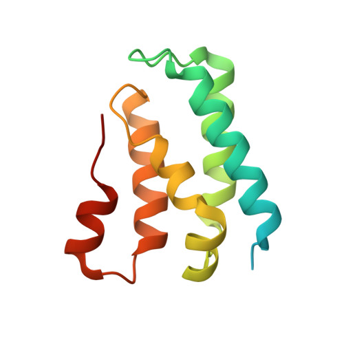
The elongation factor TFIIS interacts with Paf1C complex to facilitate processive transcription by Pol II. We here determined the crystal structure of the trypanosoma TFIIS LW domain in a complex with the LFG motif of Leo1, as well as the structures of apo-form TFIIS LW domains from trypanosoma, yeast and human. We revealed that all three TFIIS LW domains possess a conserved hydrophobic core that mediates their interactions with Leo1. Intriguingly, the structural study revealed that trypanosoma Leo1 binding induces the TFIIS LW domain to undergo a conformational change reflected in the length and orientation of α6 helix that is absent in the yeast and human counterparts. These differences explain the higher binding affinity of the TFIIS LW domain interacting with Leo1 in trypanosoma than in yeast and human, and indicate species-specific variations in the interactions. Importantly, the interactions between the TFIIS LW domain and an LFG motif of Leo1 were found to be critical for TFIIS to anchor the entire Paf1C complex. Thus, in addition to revealing a detailed structural basis for the TFIIS-Paf1C interaction, our studies also shed light on the origin and evolution of the roles of TFIIS and Paf1C complex in regulation of transcription elongation.
Organizational Affiliation:
MOE Key Laboratory for Membraneless Organelles and Cellular Dynamics, Hefei National Research Center for Interdisciplinary Sciences at the Microscale, School of Life Sciences, University of Science and Technology of China, Hefei, Anhui 230022, PR China; Department of Ophthalmology, The Second Affiliated Hospital of Anhui Medical University, 678 Furong Road, Hefei, Anhui, PR China.