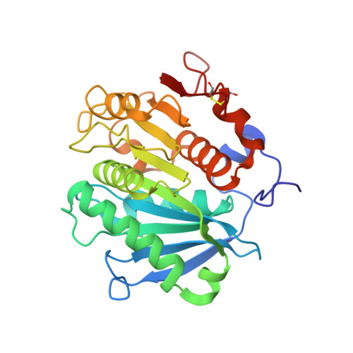
7XTT

The structure of engineered TfCut S130A in complex with MHET


Experimental Data Snapshot

  • Method: X-RAY DIFFRACTION
  • Resolution: 1.82 Å
  • R-Value Free: 0.173 
  • R-Value Work: 0.136 
  • R-Value Observed: 0.138 

Starting Model: experimental
View more details

wwPDB Validation   3D Report Full Report


Ligand Structure Quality Assessment 


This is version 1.3 of the entry. See complete history


Literature

Complete bio-degradation of poly(butylene adipate-co-terephthalate) via engineered cutinases.

Yang, Y.Min, J.Xue, T.Jiang, P.Liu, X.Peng, R.Huang, J.W.Qu, Y.Li, X.Ma, N.Tsai, F.C.Dai, L.Zhang, Q.Liu, Y.Chen, C.C.Guo, R.T.

(2023) Nat Commun 14: 1645-1645

  • DOI: https://doi.org/10.1038/s41467-023-37374-3
  • Primary Citation of Related Structures:  
    7XTR, 7XTS, 7XTT, 7XTU, 7XTV, 7XTW

  • PubMed Abstract: 

    Poly(butylene adipate-co-terephthalate) (PBAT), a polyester made of terephthalic acid (TPA), 1,4-butanediol, and adipic acid, is extensively utilized in plastic production and has accumulated globally as environmental waste. Biodegradation is an attractive strategy to manage PBAT, but an effective PBAT-degrading enzyme is required. Here, we demonstrate that cutinases are highly potent enzymes that can completely decompose PBAT films in 48 h. We further show that the engineered cutinases, by applying a double mutation strategy to render a more flexible substrate-binding pocket exhibit higher decomposition rates. Notably, these variants produce TPA as a major end-product, which is beneficial feature for the future recycling economy. The crystal structures of wild type and double mutation of a cutinase from Thermobifida fusca in complex with a substrate analogue are also solved, elucidating their substrate-binding modes. These structural and biochemical analyses enable us to propose the mechanism of cutinase-mediated PBAT degradation.


  • Organizational Affiliation

    State Key Laboratory of Biocatalysis and Enzyme Engineering, Hubei Hongshan Laboratory, Hubei Collaborative Innovation Center for Green Transformation of Bio-Resources, Hubei Key Laboratory of Industrial Biotechnology, School of Life Sciences, Hubei University, 430062, Wuhan, People's Republic of China.


Macromolecules
Find similar proteins by:  (by identity cutoff)  |  3D Structure
Entity ID: 1
MoleculeChains Sequence LengthOrganismDetailsImage
alpha/beta hydrolase
A, B
262Thermobifida fuscaMutation(s): 0 
Gene Names: cut-2.KW3
EC: 3.1.1.74
Entity Groups  
Sequence Clusters30% Identity50% Identity70% Identity90% Identity95% Identity100% Identity
Sequence Annotations
Expand
  • Reference Sequence
Small Molecules
Ligands 4 Unique
IDChains Name / Formula / InChI Key2D Diagram3D Interactions
C9C (Subject of Investigation/LOI)
Query on C9C

Download Ideal Coordinates CCD File 
D [auth B]4-(2-hydroxyethyloxycarbonyl)benzoic acid
C10 H10 O5
BCBHDSLDGBIFIX-UHFFFAOYSA-N
SO4
Query on SO4

Download Ideal Coordinates CCD File 
G [auth B]SULFATE ION
O4 S
QAOWNCQODCNURD-UHFFFAOYSA-L
EDO
Query on EDO

Download Ideal Coordinates CCD File 
C [auth A],
E [auth B],
F [auth B]
1,2-ETHANEDIOL
C2 H6 O2
LYCAIKOWRPUZTN-UHFFFAOYSA-N
NA
Query on NA

Download Ideal Coordinates CCD File 
H [auth B],
I [auth B]
SODIUM ION
Na
FKNQFGJONOIPTF-UHFFFAOYSA-N
Experimental Data & Validation

Experimental Data

  • Method: X-RAY DIFFRACTION
  • Resolution: 1.82 Å
  • R-Value Free: 0.173 
  • R-Value Work: 0.136 
  • R-Value Observed: 0.138 
  • Space Group: P 1 21 1
Unit Cell:
Length ( Å )Angle ( ˚ )
a = 65.838α = 90
b = 41.903β = 92.11
c = 80.868γ = 90
Software Package:
Software NamePurpose
SADABSdata scaling
PHENIXrefinement
PDB_EXTRACTdata extraction
SAINTdata reduction
PHASERphasing

Structure Validation

View Full Validation Report


Ligand Structure Quality Assessment 


Entry History & Funding Information

Deposition Data


Funding OrganizationLocationGrant Number
National Natural Science Foundation of China (NSFC)China31870790
National Natural Science Foundation of China (NSFC)China31971205
National Natural Science Foundation of China (NSFC)China32101016

Revision History  (Full details and data files)

  • Version 1.0: 2023-03-29
    Type: Initial release
  • Version 1.1: 2023-04-12
    Changes: Database references
  • Version 1.2: 2023-11-29
    Changes: Data collection, Refinement description
  • Version 1.3: 2024-11-20
    Changes: Structure summary