7VQ0

Cryo-EM structure of the SARS-CoV-2 spike protein (2-up RBD) bound to neutralizing nanobodies P86


Experimental Data Snapshot

  • Method: ELECTRON MICROSCOPY
  • Resolution: 3.03 Å
  • Aggregation State: PARTICLE 
  • Reconstruction Method: SINGLE PARTICLE 

wwPDB Validation   3D Report Full Report


This is version 1.0 of the entry. See complete history


Literature

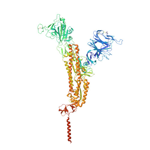
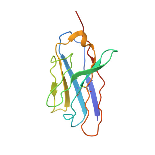
A panel of nanobodies recognizing conserved hidden clefts of all SARS-CoV-2 spike variants including Omicron.

Maeda, R.Fujita, J.Konishi, Y.Kazuma, Y.Yamazaki, H.Anzai, I.Watanabe, T.Yamaguchi, K.Kasai, K.Nagata, K.Yamaoka, Y.Miyakawa, K.Ryo, A.Shirakawa, K.Sato, K.Makino, F.Matsuura, Y.Inoue, T.Imura, A.Namba, K.Takaori-Kondo, A.

(2022) Commun Biol 5: 669-669

  • DOI: https://doi.org/10.1038/s42003-022-03630-3
  • Primary Citation of Related Structures:  
    7VPY, 7VQ0

  • PubMed Abstract: 

    We are amid the historic coronavirus infectious disease 2019 (COVID-19) pandemic. Imbalances in the accessibility of vaccines, medicines, and diagnostics among countries, regions, and populations, and those in war crises, have been problematic. Nanobodies are small, stable, customizable, and inexpensive to produce. Herein, we present a panel of nanobodies that can detect the spike proteins of five SARS-CoV-2 variants of concern (VOCs) including Omicron. Here we show via ELISA, lateral flow, kinetic, flow cytometric, microscopy, and Western blotting assays that our nanobodies can quantify the spike variants. This panel of nanobodies broadly neutralizes viral infection caused by pseudotyped and authentic SARS-CoV-2 VOCs. Structural analyses show that the P86 clone targets epitopes that are conserved yet unclassified on the receptor-binding domain (RBD) and contacts the N-terminal domain (NTD). Human antibodies rarely access both regions; consequently, the clone buries hidden crevasses of SARS-CoV-2 spike proteins that go undetected by conventional antibodies.


  • Organizational Affiliation

    Department of Haematology and Oncology, Graduate School of Medicine, Kyoto University, Kyoto, 606-8507, Japan. maeda@cognano.co.jp.


Macromolecules
Find similar proteins by:  (by identity cutoff)  |  3D Structure
Entity ID: 1
MoleculeChains Sequence LengthOrganismDetailsImage
Spike glycoprotein
A, B, C
1,247Severe acute respiratory syndrome coronavirus 2Mutation(s): 6 
Gene Names: S2
UniProt
Find proteins for P0DTC2 (Severe acute respiratory syndrome coronavirus 2)
Explore P0DTC2 
Go to UniProtKB:  P0DTC2
Entity Groups  
Sequence Clusters30% Identity50% Identity70% Identity90% Identity95% Identity100% Identity
UniProt GroupP0DTC2
Sequence Annotations
Expand
  • Reference Sequence
Find similar proteins by:  (by identity cutoff)  |  3D Structure
Entity ID: 2
MoleculeChains Sequence LengthOrganismDetailsImage
Neutralizing nanobody P86
D, E, F
122Vicugna pacosMutation(s): 0 
Entity Groups  
Sequence Clusters30% Identity50% Identity70% Identity90% Identity95% Identity100% Identity
Sequence Annotations
Expand
  • Reference Sequence
Oligosaccharides

Help

Entity ID: 4
MoleculeChains Length2D Diagram Glycosylation3D Interactions
2-acetamido-2-deoxy-beta-D-glucopyranose-(1-4)-[alpha-L-fucopyranose-(1-6)]2-acetamido-2-deoxy-beta-D-glucopyranoseDA [auth d],
N,
V
3N/AN-Glycosylation
Glycosylation Resources
GlyTouCan:  G21290RB
GlyCosmos:  G21290RB
GlyGen:  G21290RB
Entity ID: 5
MoleculeChains Length2D Diagram Glycosylation3D Interactions
alpha-D-mannopyranose-(1-4)-2-acetamido-2-deoxy-beta-D-glucopyranose-(1-4)-2-acetamido-2-deoxy-beta-D-glucopyranose
EA [auth e],
FA [auth f],
O,
P,
W,
EA [auth e],
FA [auth f],
O,
P,
W,
X
3N/AN-Glycosylation
Glycosylation Resources
GlyTouCan:  G62182OO
GlyCosmos:  G62182OO
GlyGen:  G62182OO
Small Molecules
Ligands 1 Unique
IDChains Name / Formula / InChI Key2D Diagram3D Interactions
NAG
Query on NAG

Download Ideal Coordinates CCD File 
AB [auth C]
BB [auth C]
CB [auth C]
DB [auth C]
EB [auth C]
AB [auth C],
BB [auth C],
CB [auth C],
DB [auth C],
EB [auth C],
FB [auth C],
GA [auth A],
GB [auth C],
HA [auth A],
HB [auth C],
IA [auth A],
IB [auth C],
JA [auth A],
JB [auth C],
KA [auth A],
LA [auth A],
MA [auth A],
NA [auth A],
OA [auth A],
PA [auth A],
QA [auth B],
RA [auth B],
SA [auth B],
TA [auth B],
UA [auth B],
VA [auth B],
WA [auth B],
XA [auth B],
YA [auth B],
ZA [auth C]
2-acetamido-2-deoxy-beta-D-glucopyranose
C8 H15 N O6
OVRNDRQMDRJTHS-FMDGEEDCSA-N
Experimental Data & Validation

Experimental Data

  • Method: ELECTRON MICROSCOPY
  • Resolution: 3.03 Å
  • Aggregation State: PARTICLE 
  • Reconstruction Method: SINGLE PARTICLE 
EM Software:
TaskSoftware PackageVersion
RECONSTRUCTIONcryoSPARC3.2.0
MODEL REFINEMENTPHENIX1.19.1-4122

Structure Validation

View Full Validation Report


Entry History & Funding Information

Deposition Data


Funding OrganizationLocationGrant Number
Japan Agency for Medical Research and Development (AMED)JapanJP20fk0108268
Japan Agency for Medical Research and Development (AMED)JapanJP20fk018517
Japan Agency for Medical Research and Development (AMED)JapanJP20fk018413
Japan Society for the Promotion of Science (JSPS)JapanJP20K22630
Japan Society for the Promotion of Science (JSPS)JapanJP25000013
Japan Agency for Medical Research and Development (AMED)JapanJP21am0101117
Japan Agency for Medical Research and Development (AMED)JapanJP17pc0101020
Japan Science and TechnologyJapanJPMJOP1861

Revision History  (Full details and data files)

  • Version 1.0: 2022-07-20
    Type: Initial release