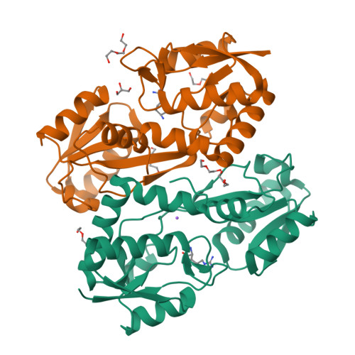
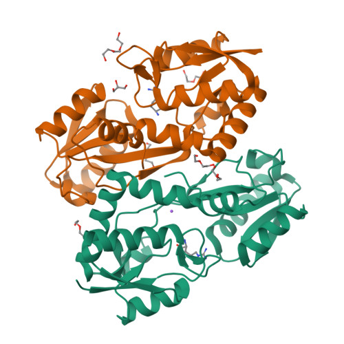
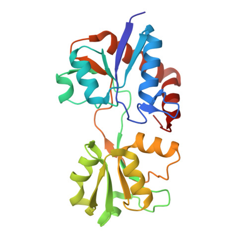
8B5E

Exploring the ligand binding and conformational dynamics of receptor domain 1 of the ABC transporter GlnPQ


Experimental Data Snapshot

  • Method: X-RAY DIFFRACTION
  • Resolution: 1.60 Å
  • R-Value Free: 0.200 
  • R-Value Work: 0.100 

wwPDB Validation   3D Report Full Report


Ligand Structure Quality Assessment 


This is version 1.1 of the entry. See complete history


Literature

Exploring the Ligand Binding and Conformational Dynamics of the Substrate-Binding Domain 1 of the ABC Transporter GlnPQ.

Nemchinova, M.Schuurman-Wolters, G.K.Whittaker, J.J.Arkhipova, V.Marrink, S.J.Poolman, B.Guskov, A.

(2024) J Phys Chem B 128: 7822-7832

  • DOI: https://doi.org/10.1021/acs.jpcb.4c02662
  • Primary Citation of Related Structures:  
    8B5D, 8B5E

  • PubMed Abstract: 

    The adenosine triphosphate (ATP)-binding cassette (ABC) importer GlnPQ from Lactococcus lactis has two sequential covalently linked substrate-binding domains (SBDs), which capture the substrates and deliver them to the translocon. The two SBDs differ in their ligand specificities, binding affinities and the distance to the transmembrane domain; interestingly, both SBDs can bind their ligands simultaneously without affecting each other. In this work, we studied the binding of ligands to both SBDs using X-ray crystallography and molecular dynamics simulations. We report three high-resolution structures of SBD1, namely, the wild-type SBD1 with bound asparagine or arginine, and E184D SBD1 with glutamine bound. Molecular dynamics (MD) simulations provide a detailed insight into the dynamics associated with open-closed transitions of the SBDs.


  • Organizational Affiliation

    Groningen Institute for Biomolecular Sciences and Biotechnology, University of Groningen, 9747AG Groningen, The Netherlands.


Macromolecules
Find similar proteins by:  (by identity cutoff)  |  3D Structure
Entity ID: 1
MoleculeChains Sequence LengthOrganismDetailsImage
ABC transporter permease subunit
A, B
225Lactococcus lactisMutation(s): 0 
Gene Names: glnPAMHIJAGA_02474GII02_01815
UniProt
Find proteins for A0A2X0R690 (Lactococcus lactis)
Explore A0A2X0R690 
Go to UniProtKB:  A0A2X0R690
Entity Groups  
Sequence Clusters30% Identity50% Identity70% Identity90% Identity95% Identity100% Identity
UniProt GroupA0A2X0R690
Sequence Annotations
Expand
  • Reference Sequence
Small Molecules
Ligands 5 Unique
IDChains Name / Formula / InChI Key2D Diagram3D Interactions
ARG (Subject of Investigation/LOI)
Query on ARG

Download Ideal Coordinates CCD File 
C [auth A],
F [auth B]
ARGININE
C6 H15 N4 O2
ODKSFYDXXFIFQN-BYPYZUCNSA-O
PGE
Query on PGE

Download Ideal Coordinates CCD File 
L [auth B]TRIETHYLENE GLYCOL
C6 H14 O4
ZIBGPFATKBEMQZ-UHFFFAOYSA-N
PEG
Query on PEG

Download Ideal Coordinates CCD File 
G [auth B],
H [auth B],
I [auth B],
J [auth B],
K [auth B]
DI(HYDROXYETHYL)ETHER
C4 H10 O3
MTHSVFCYNBDYFN-UHFFFAOYSA-N
GOL
Query on GOL

Download Ideal Coordinates CCD File 
E [auth B]GLYCEROL
C3 H8 O3
PEDCQBHIVMGVHV-UHFFFAOYSA-N
NA
Query on NA

Download Ideal Coordinates CCD File 
D [auth A]SODIUM ION
Na
FKNQFGJONOIPTF-UHFFFAOYSA-N
Experimental Data & Validation

Experimental Data

  • Method: X-RAY DIFFRACTION
  • Resolution: 1.60 Å
  • R-Value Free: 0.200 
  • R-Value Work: 0.100 
  • Space Group: P 1
Unit Cell:
Length ( Å )Angle ( ˚ )
a = 34.67α = 92.4
b = 53.08β = 92.37
c = 54.08γ = 94.11
Software Package:
Software NamePurpose
REFMACrefinement
XDSdata reduction
Aimlessdata scaling
PHASERphasing

Structure Validation

View Full Validation Report


Ligand Structure Quality Assessment 


Entry History & Funding Information

Deposition Data


Funding OrganizationLocationGrant Number
Not funded--

Revision History  (Full details and data files)

  • Version 1.0: 2023-10-04
    Type: Initial release
  • Version 1.1: 2024-09-04
    Changes: Database references