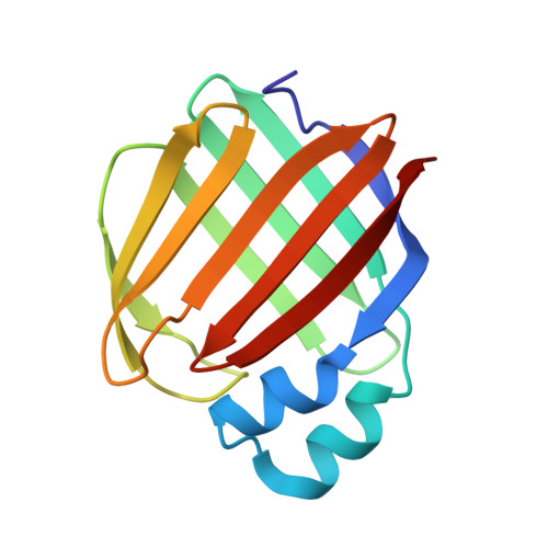
8D6N

Q108K:K40L:T53A:R58L:Q38F:Q4F mutant of hCRBPII bound to synthetic fluorophore CM1V


Experimental Data Snapshot

  • Method: X-RAY DIFFRACTION
  • Resolution: 1.42 Å
  • R-Value Free: 0.233 
  • R-Value Work: 0.213 
  • R-Value Observed: 0.214 

Starting Model: experimental
View more details

wwPDB Validation   3D Report Full Report


Ligand Structure Quality Assessment 


This is version 1.4 of the entry. See complete history


Literature

Light controlled reversible Michael addition of cysteine: a new tool for dynamic site-specific labeling of proteins.

Maity, S.Bingham, C.Sheng, W.Ehyaei, N.Chakraborty, D.Tahmasebi-Nick, S.Kimmel, T.E.Vasileiou, C.Geiger, J.H.Borhan, B.

(2023) Analyst 148: 1085-1092

  • DOI: https://doi.org/10.1039/d2an01395a
  • Primary Citation of Related Structures:  
    8D6H, 8D6L, 8D6N, 8DB2, 8DN1

  • PubMed Abstract: 

    Cysteine-based Michael addition is a widely employed strategy for covalent conjugation of proteins, peptides, and drugs. The covalent reaction is irreversible in most cases, leading to a lack of control over the process. Utilizing spectroscopic analyses along with X-ray crystallographic studies, we demonstrate Michael addition of an engineered cysteine residue in human Cellular Retinol Binding Protein II (hCRBPII) with a coumarin analog that creates a non-fluorescent complex. UV-illumination reverses the conjugation, yielding a fluorescent species, presumably through a retro -Michael process. This series of events can be repeated between a bound and non-bound form of the cysteine reversibly, resulting in the ON-OFF control of fluorescence. The details of the mechanism of photoswitching was illuminated by recapitulation of the process in light irradiated single crystals, confirming the mechanism at atomic resolution.


  • Organizational Affiliation

    Department of Chemistry, Michigan State University, 578 S. Shaw Ln., East Lansing, MI 48824, USA. babak@chemistry.msu.edu.


Macromolecules
Find similar proteins by:  (by identity cutoff)  |  3D Structure
Entity ID: 1
MoleculeChains Sequence LengthOrganismDetailsImage
Retinol-binding protein 2133Homo sapiensMutation(s): 6 
Gene Names: RBP2CRBP2
UniProt & NIH Common Fund Data Resources
Find proteins for P50120 (Homo sapiens)
Explore P50120 
Go to UniProtKB:  P50120
PHAROS:  P50120
GTEx:  ENSG00000114113 
Entity Groups  
Sequence Clusters30% Identity50% Identity70% Identity90% Identity95% Identity100% Identity
UniProt GroupP50120
Sequence Annotations
Expand
  • Reference Sequence
Small Molecules
Ligands 4 Unique
IDChains Name / Formula / InChI Key2D Diagram3D Interactions
RH6
Query on RH6

Download Ideal Coordinates CCD File 
C [auth A](2E)-3-[7-(diethylamino)-2-oxo-2H-1-benzopyran-3-yl]prop-2-enal, bound form
C16 H19 N O2
YRIYSICZBXXPLJ-QPJJXVBHSA-N
MLA
Query on MLA

Download Ideal Coordinates CCD File 
L [auth A],
M [auth A]
MALONIC ACID
C3 H4 O4
OFOBLEOULBTSOW-UHFFFAOYSA-N
GOL
Query on GOL

Download Ideal Coordinates CCD File 
D [auth A]
F [auth A]
H [auth A]
I [auth A]
J [auth A]
D [auth A],
F [auth A],
H [auth A],
I [auth A],
J [auth A],
K [auth A],
N [auth A]
GLYCEROL
C3 H8 O3
PEDCQBHIVMGVHV-UHFFFAOYSA-N
ACT
Query on ACT

Download Ideal Coordinates CCD File 
B [auth A],
E [auth A],
G [auth A]
ACETATE ION
C2 H3 O2
QTBSBXVTEAMEQO-UHFFFAOYSA-M
Experimental Data & Validation

Experimental Data

  • Method: X-RAY DIFFRACTION
  • Resolution: 1.42 Å
  • R-Value Free: 0.233 
  • R-Value Work: 0.213 
  • R-Value Observed: 0.214 
  • Space Group: C 1 2 1
Unit Cell:
Length ( Å )Angle ( ˚ )
a = 29.612α = 90
b = 67.106β = 90.108
c = 63.903γ = 90
Software Package:
Software NamePurpose
PHENIXrefinement
HKL-2000data reduction
HKL-2000data scaling
PHASERphasing

Structure Validation

View Full Validation Report


Ligand Structure Quality Assessment 


Entry History & Funding Information

Deposition Data


Funding OrganizationLocationGrant Number
National Institutes of Health/National Institute of Biomedical Imaging and Bioengineering (NIH/NIBIB)United States--

Revision History  (Full details and data files)

  • Version 1.0: 2023-02-01
    Type: Initial release
  • Version 1.1: 2023-02-22
    Changes: Database references
  • Version 1.2: 2023-03-15
    Changes: Database references
  • Version 1.3: 2023-10-25
    Changes: Data collection, Refinement description
  • Version 1.4: 2024-10-16
    Changes: Structure summary