8DWR | pdb_00008dwr

Crystal structure of the L333V variant of catalase-peroxidase from Mycobacterium tuberculosis


Experimental Data Snapshot

  • Method: X-RAY DIFFRACTION
  • Resolution: 2.10 Å
  • R-Value Free: 
    0.227 (Depositor), 0.230 (DCC) 
  • R-Value Work: 
    0.181 (Depositor), 0.180 (DCC) 
  • R-Value Observed: 
    0.181 (Depositor) 

wwPDB Validation   3D Report Full Report


Ligand Structure Quality Assessment 

Created with Raphaël 2.3.0Worse 01 BetterLigand structure goodness of fit to experimental dataBest fitted HEMClick on this verticalbar to view details

This is version 1.1 of the entry. See complete history


Literature

Characterization of a catalase-peroxidase variant (L333V-KatG) identified in an INH-resistant Mycobacterium tuberculosis clinical isolate.

Uribe-Vazquez, B.Diaz-Vilchis, A.Avila-Linares, A.Saab-Rincon, G.Marin-Tovar, Y.Flores, H.Pastor, N.Huerta-Miranda, G.Rudino-Pinera, E.Soberon, X.

(2024) Biochem Biophys Rep 37: 101649-101649

  • DOI: https://doi.org/10.1016/j.bbrep.2024.101649
  • Primary Citation of Related Structures:  
    8DWR

  • PubMed Abstract: 

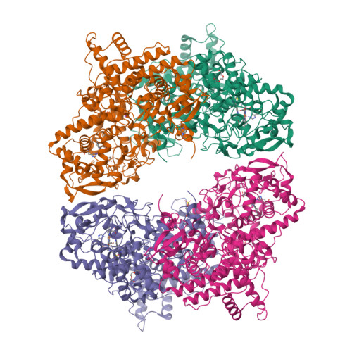
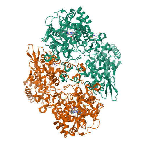
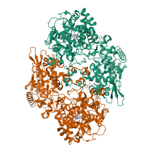
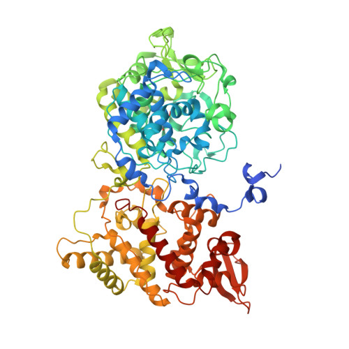
    Mycobacterium tuberculosis catalase-peroxidase ( Mt -KatG) is a bifunctional heme-dependent enzyme that has been shown to activate isoniazid (INH), the widely used antibiotic against tuberculosis (TB). The L333V-KatG variant has been associated with INH resistance in clinical M. tuberculosis isolates from Mexico. To understand better the mechanisms of INH activation, its catalytic properties (catalase, peroxidase, and IN-NAD formation) and crystal structure were compared with those of the wild-type enzyme (WT-KatG). The rate of IN-NAD formation mediated by WT-KatG was 23% greater than L333V-KatG when INH concentration is varied. In contrast to WT-KatG, the crystal structure of the L333V-KatG variant has a perhydroxy modification of the indole nitrogen of W107 from MYW adduct. L333V-KatG shows most of the active site residues in a similar position to WT-KatG; only R418 is in the R-conformation instead of the double R and Y conformation present in WT-KatG. L333V-KatG shows a small displacement respect to WT-KatG in the helix from R385 to L404 towards the mutation site, an increase in length of the coordination bond between H270 and heme Fe, and a longer H-bond between proximal D381 and W321, compared to WT-KatG; these small displacements could explain the altered redox potential of the heme, and result in a less active and stable enzyme.


  • Organizational Affiliation

    Departamento de Ingeniería Celular y Biocatálisis, Instituto de Biotecnología, UNAM, Avenida Universidad 2001, Colonia Chamilpa, 62210, Cuernavaca, México.


Macromolecules
Find similar proteins by:  (by identity cutoff)  |  3D Structure
Entity ID: 1
MoleculeChains Sequence LengthOrganismDetailsImage
Catalase-peroxidase
A, B, C, D
746Mycobacterium tuberculosisMutation(s): 1 
Gene Names: katGRv1908cMTCY180.10
EC: 1.11.1.21
UniProt
Find proteins for P9WIE5 (Mycobacterium tuberculosis (strain ATCC 25618 / H37Rv))
Explore P9WIE5 
Go to UniProtKB:  P9WIE5
Entity Groups  
Sequence Clusters30% Identity50% Identity70% Identity90% Identity95% Identity100% Identity
UniProt GroupP9WIE5
Sequence Annotations
Expand
  • Reference Sequence
Small Molecules
Ligands 4 Unique
IDChains Name / Formula / InChI Key2D Diagram3D Interactions
HEM (Subject of Investigation/LOI)
Query on HEM

Download Ideal Coordinates CCD File 
E [auth A],
J [auth B],
M [auth C],
R [auth D]
PROTOPORPHYRIN IX CONTAINING FE
C34 H32 Fe N4 O4
KABFMIBPWCXCRK-RGGAHWMASA-L
EPE
Query on EPE

Download Ideal Coordinates CCD File 
F [auth A],
I [auth B],
N [auth C],
P [auth C]
4-(2-HYDROXYETHYL)-1-PIPERAZINE ETHANESULFONIC ACID
C8 H18 N2 O4 S
JKMHFZQWWAIEOD-UHFFFAOYSA-N
PGE
Query on PGE

Download Ideal Coordinates CCD File 
H [auth A],
L [auth B],
Q [auth C],
T [auth D]
TRIETHYLENE GLYCOL
C6 H14 O4
ZIBGPFATKBEMQZ-UHFFFAOYSA-N
NA
Query on NA

Download Ideal Coordinates CCD File 
G [auth A],
K [auth B],
O [auth C],
S [auth D]
SODIUM ION
Na
FKNQFGJONOIPTF-UHFFFAOYSA-N
Modified Residues  1 Unique
IDChains TypeFormula2D DiagramParent
TOX
Query on TOX
A, B, C, D
L-PEPTIDE LINKINGC11 H12 N2 O4TRP
Experimental Data & Validation

Experimental Data

  • Method: X-RAY DIFFRACTION
  • Resolution: 2.10 Å
  • R-Value Free:  0.227 (Depositor), 0.230 (DCC) 
  • R-Value Work:  0.181 (Depositor), 0.180 (DCC) 
  • R-Value Observed: 0.181 (Depositor) 
Space Group: P 1 21 1
Unit Cell:
Length ( Å )Angle ( ˚ )
a = 82.671α = 90
b = 150.817β = 90.45
c = 127.037γ = 90
Software Package:
Software NamePurpose
PHENIXrefinement
PDB_EXTRACTdata extraction
XDSdata reduction
XDSdata scaling
PHASERphasing

Structure Validation

View Full Validation Report


Ligand Structure Quality Assessment 

Created with Raphaël 2.3.0Worse 01 BetterLigand structure goodness of fit to experimental dataBest fitted HEMClick on this verticalbar to view details

Entry History & Funding Information

Deposition Data


Funding OrganizationLocationGrant Number
Other governmentMexicoIBt, UNAM Institutional budget

Revision History  (Full details and data files)

  • Version 1.0: 2023-08-02
    Type: Initial release
  • Version 1.1: 2024-03-13
    Changes: Data collection, Database references