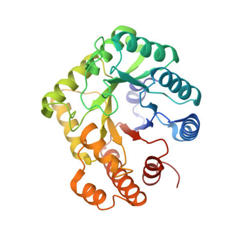
5-Formyltetrahydrofolate promotes conformational remodeling in a methylenetetrahydrofolate reductase active site and inhibits its activity.
Yamada, K., Mendoza, J., Koutmos, M.(2022) J Biol Chem 299: 102855-102855
- PubMed: 36592927
- DOI: https://doi.org/10.1016/j.jbc.2022.102855
- Primary Citation of Related Structures:
7TH4, 7TH5, 8EAC - PubMed Abstract:
The flavoprotein methylenetetrahydrofolate reductase (MTHFR) catalyzes the reduction of N5, N10-methylenetetrahydrofolate (CH 2 -H 4 folate) to N5-methyltetrahydrofolate (CH 3 -H 4 folate), committing a methyl group from the folate cycle to the methionine one. This committed step is the sum of multiple ping-pong electron transfers involving multiple substrates, intermediates, and products all sharing the same active site. Insight into folate substrate binding is needed to better understand this multifunctional active site. Here, we performed activity assays with Thermus thermophilus MTHFR (tMTHFR), which showed pH-dependent inhibition by the substrate analog, N5-formyltetrahydrofolate (CHO-H 4 folate). Our crystal structure of a tMTHFR•CHO-H 4 folate complex revealed a unique folate-binding mode; tMTHFR subtly rearranges its active site to form a distinct folate-binding environment. Formation of a novel binding pocket for the CHO-H 4 folate p-aminobenzoic acid moiety directly affects how bent the folate ligand is and its accommodation in the active site. Comparative analysis of the available active (FAD- and folate-bound) MTHFR complex structures reveals that CHO-H 4 folate is accommodated in the active site in a conformation that would not support hydride transfer, but rather in a conformation that potentially reports on a different step in the reaction mechanism after this committed step, such as CH 2 -H 4 folate ring-opening. This active site remodeling provides insights into the functional relevance of the differential folate-binding modes and their potential roles in the catalytic cycle. The conformational flexibility displayed by tMTHFR demonstrates how a shared active site can use a few amino acid residues in lieu of extra domains to accommodate chemically distinct moieties and functionalities.
Organizational Affiliation:
Department of Chemistry, University of Michigan, Ann Arbor, Michigan, USA; Program in Biophysics, University of Michigan, Ann Arbor, Michigan, USA. Electronic address: yamadak@umich.edu.


