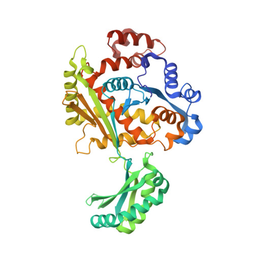
8GDN

Structure of PmHMGR bound to mevalonate, CoA and NAD.


Experimental Data Snapshot

  • Method: X-RAY DIFFRACTION
  • Resolution: 1.99 Å
  • R-Value Free: 0.225 
  • R-Value Work: 0.181 
  • R-Value Observed: 0.183 

Starting Model: experimental
View more details

wwPDB Validation   3D Report Full Report


Ligand Structure Quality Assessment 


This is version 1.0 of the entry. See complete history


Literature

pH-dependent reaction triggering in PmHMGR crystals for time-resolved crystallography.

Purohit, V.Steussy, C.N.Rosales, A.R.Critchelow, C.J.Schmidt, T.Helquist, P.Wiest, O.Mesecar, A.Cohen, A.E.Stauffacher, C.V.

(2024) Biophys J 123: 622-637

  • DOI: https://doi.org/10.1016/j.bpj.2024.02.003
  • Primary Citation of Related Structures:  
    8GDN, 8SZ6, 8VLQ

  • PubMed Abstract: 

    Serial crystallography and time-resolved data collection can readily be employed to investigate the catalytic mechanism of Pseudomonas mevalonii 3-hydroxy-3-methylglutaryl (HMG)-coenzyme-A (CoA) reductase (PmHMGR) by changing the environmental conditions in the crystal and so manipulating the reaction rate. This enzyme uses a complex mechanism to convert mevalonate to HMG-CoA using the co-substrate CoA and cofactor NAD + . The multi-step reaction mechanism involves an exchange of bound NAD + and large conformational changes by a 50-residue subdomain. The enzymatic reaction can be run in both forward and reverse directions in solution and is catalytically active in the crystal for multiple reaction steps. Initially, the enzyme was found to be inactive in the crystal starting with bound mevalonate, CoA, and NAD + . To observe the reaction from this direction, we examined the effects of crystallization buffer constituents and pH on enzyme turnover, discovering a strong inhibition in the crystallization buffer and a controllable increase in enzyme turnover as a function of pH. The inhibition is dependent on ionic concentration of the crystallization precipitant ammonium sulfate but independent of its ionic composition. Crystallographic studies show that the observed inhibition only affects the oxidation of mevalonate but not the subsequent reactions of the intermediate mevaldehyde. Calculations of the pKa values for the enzyme active site residues suggest that the effect of pH on turnover is due to the changing protonation state of His381. We have now exploited the changes in ionic inhibition in combination with the pH-dependent increase in turnover as a novel approach for triggering the PmHMGR reaction in crystals and capturing information about its intermediate states along the reaction pathway.


  • Organizational Affiliation

    Department of Biological Sciences, Purdue University, West Lafayette, Indiana.


Macromolecules
Find similar proteins by:  (by identity cutoff)  |  3D Structure
Entity ID: 1
MoleculeChains Sequence LengthOrganismDetailsImage
3-hydroxy-3-methylglutaryl-coenzyme A reductase
A, B
428Pseudomonas sp. 'mevaloniiMutation(s): 0 
Gene Names: mvaA
EC: 1.1.1.88
UniProt
Find proteins for P13702 (Pseudomonas mevalonii)
Explore P13702 
Go to UniProtKB:  P13702
Entity Groups  
Sequence Clusters30% Identity50% Identity70% Identity90% Identity95% Identity100% Identity
UniProt GroupP13702
Sequence Annotations
Expand
  • Reference Sequence
Small Molecules
Ligands 4 Unique
IDChains Name / Formula / InChI Key2D Diagram3D Interactions
COA (Subject of Investigation/LOI)
Query on COA

Download Ideal Coordinates CCD File 
C [auth A]COENZYME A
C21 H36 N7 O16 P3 S
RGJOEKWQDUBAIZ-IBOSZNHHSA-N
NAD (Subject of Investigation/LOI)
Query on NAD

Download Ideal Coordinates CCD File 
G [auth B]NICOTINAMIDE-ADENINE-DINUCLEOTIDE
C21 H27 N7 O14 P2
BAWFJGJZGIEFAR-NNYOXOHSSA-N
MEV (Subject of Investigation/LOI)
Query on MEV

Download Ideal Coordinates CCD File 
D [auth A],
H [auth B]
(R)-MEVALONATE
C6 H11 O4
KJTLQQUUPVSXIM-ZCFIWIBFSA-M
SO4
Query on SO4

Download Ideal Coordinates CCD File 
E [auth A],
F [auth A]
SULFATE ION
O4 S
QAOWNCQODCNURD-UHFFFAOYSA-L
Experimental Data & Validation

Experimental Data

  • Method: X-RAY DIFFRACTION
  • Resolution: 1.99 Å
  • R-Value Free: 0.225 
  • R-Value Work: 0.181 
  • R-Value Observed: 0.183 
  • Space Group: I 41 3 2
Unit Cell:
Length ( Å )Angle ( ˚ )
a = 226.043α = 90
b = 226.043β = 90
c = 226.043γ = 90
Software Package:
Software NamePurpose
PHENIXrefinement
HKL-2000data reduction
HKL-2000data scaling
PDB_EXTRACTdata extraction
PHASERphasing

Structure Validation

View Full Validation Report


Ligand Structure Quality Assessment 


Entry History & Funding Information

Deposition Data


Funding OrganizationLocationGrant Number
National Institutes of Health/National Institute of General Medical Sciences (NIH/NIGMS)United StatesRO1 GM111645
National Institutes of Health/National Cancer Institute (NIH/NCI)United StatesP30 CA023168

Revision History  (Full details and data files)

  • Version 1.0: 2024-03-20
    Type: Initial release