8IXL

top segment of the bacteriophage M13 mini variant

  • Classification: VIRAL PROTEIN
  • Organism(s): Inovirus M13
  • Expression System: Escherichia coli
  • Mutation(s): No 

  • Deposited: 2023-04-01 Released: 2023-09-13 
  • Deposition Author(s): Xiang, Y., Jia, Q.
  • Funding Organization(s): Ministry of Science and Technology (MoST, China), National Natural Science Foundation of China (NSFC)

Experimental Data Snapshot

  • Method: ELECTRON MICROSCOPY
  • Resolution: 3.50 Å
  • Aggregation State: PARTICLE 
  • Reconstruction Method: SINGLE PARTICLE 

wwPDB Validation   3D Report Full Report


This is version 1.1 of the entry. See complete history


Literature

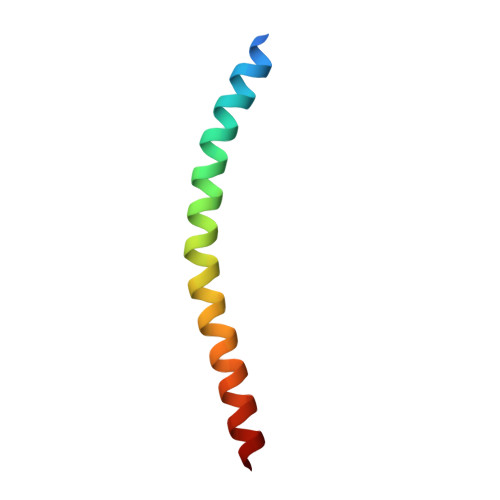
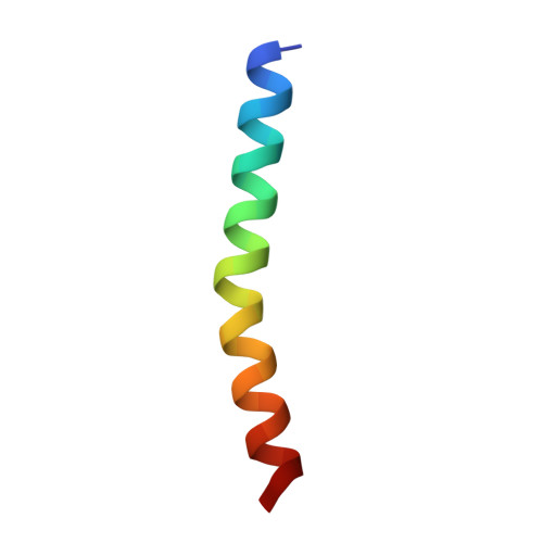
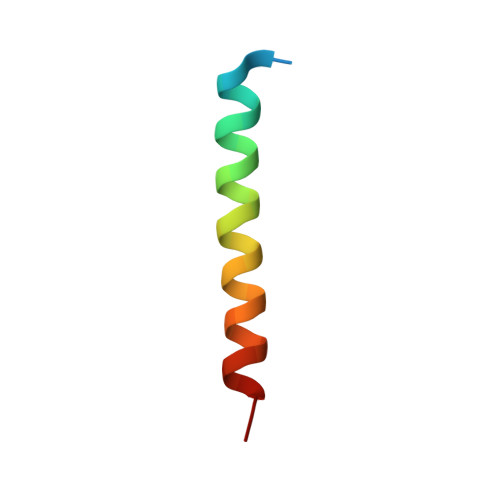
Cryo-EM structure of a bacteriophage M13 mini variant.

Jia, Q.Xiang, Y.

(2023) Nat Commun 14: 5421-5421

  • DOI: https://doi.org/10.1038/s41467-023-41151-7
  • Primary Citation of Related Structures:  
    8IXJ, 8IXK, 8IXL, 8JWT

  • PubMed Abstract: 
  • Filamentous bacteriophages package their circular, single stranded DNA genome with the major coat protein pVIII and the minor coat proteins pIII, pVII, pVI, and pIX. Here, we report the cryo-EM structure of a ~500 Å long bacteriophage M13 mini variant. The distal ends of the mini phage are sealed by two cap-like complexes composed of the minor coat proteins ...

    Filamentous bacteriophages package their circular, single stranded DNA genome with the major coat protein pVIII and the minor coat proteins pIII, pVII, pVI, and pIX. Here, we report the cryo-EM structure of a ~500 Å long bacteriophage M13 mini variant. The distal ends of the mini phage are sealed by two cap-like complexes composed of the minor coat proteins. The top cap complex consists of pVII and pIX, both exhibiting a single helix structure. Arg33 of pVII and Glu29 of pIX, located on the inner surface of the cap, play a key role in recognizing the genome packaging signal. The bottom cap complex is formed by the hook-like structures of pIII and pVI, arranged in helix barrels. Most of the inner ssDNA genome adopts a double helix structure with a similar pitch to that of the A-form double-stranded DNA. These findings provide insights into the assembly of filamentous bacteriophages.


    Organizational Affiliation

    Beijing Frontier Research Center for Biological Structure, Center for Infectious Disease Research, SXMU-Tsinghua Collaborative Innovation Center for Frontier Medicine, Department of Basic Medical Sciences, School of Medicine, Tsinghua University, Beijing, 100084, P.R. China. yxiang@mail.tsinghua.edu.cn.


Macromolecules
Find similar proteins by:  (by identity cutoff)  |  3D Structure
Entity ID: 1
MoleculeChainsSequence LengthOrganismDetailsImage
Capsid protein G8P50Inovirus M13Mutation(s): 0 
Gene Names: VIII
UniProt
Find proteins for P69541 (Enterobacteria phage M13)
Explore P69541 
Go to UniProtKB:  P69541
Entity Groups  
Sequence Clusters30% Identity50% Identity70% Identity90% Identity95% Identity100% Identity
UniProt GroupP69541
Protein Feature View
Expand
  • Reference Sequence
Find similar proteins by:  (by identity cutoff)  |  3D Structure
Entity ID: 2
MoleculeChainsSequence LengthOrganismDetailsImage
Tail virion protein G9PD,
FA [auth GA],
K [auth J],
R [auth Q],
Y
32Inovirus M13Mutation(s): 0 
Gene Names: IX
UniProt
Find proteins for P69538 (Enterobacteria phage M13)
Explore P69538 
Go to UniProtKB:  P69538
Entity Groups  
Sequence Clusters30% Identity50% Identity70% Identity90% Identity95% Identity100% Identity
UniProt GroupP69538
Protein Feature View
Expand
  • Reference Sequence
Find similar proteins by:  (by identity cutoff)  |  3D Structure
Entity ID: 3
MoleculeChainsSequence LengthOrganismDetailsImage
Tail virion protein G7PAA [auth BA],
F,
HA [auth IA],
M [auth L],
T [auth S]
33Inovirus M13Mutation(s): 0 
Gene Names: VII
UniProt
Find proteins for P69535 (Enterobacteria phage M13)
Explore P69535 
Go to UniProtKB:  P69535
Entity Groups  
Sequence Clusters30% Identity50% Identity70% Identity90% Identity95% Identity100% Identity
UniProt GroupP69535
Protein Feature View
Expand
  • Reference Sequence
Experimental Data & Validation

Experimental Data

  • Method: ELECTRON MICROSCOPY
  • Resolution: 3.50 Å
  • Aggregation State: PARTICLE 
  • Reconstruction Method: SINGLE PARTICLE 

Structure Validation

View Full Validation Report


Entry History & Funding Information

Deposition Data


Funding OrganizationLocationGrant Number
Ministry of Science and Technology (MoST, China)China--
National Natural Science Foundation of China (NSFC)China--

Revision History  (Full details and data files)

  • Version 1.0: 2023-09-13
    Type: Initial release
  • Version 1.1: 2023-09-20
    Changes: Database references