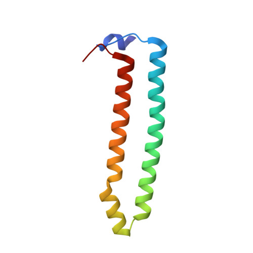
8IY0

Structure of Acb2 complexed with cAAA


Experimental Data Snapshot

  • Method: X-RAY DIFFRACTION
  • Resolution: 2.26 Å
  • R-Value Free: 0.258 
  • R-Value Work: 0.215 
  • R-Value Observed: 0.217 

Starting Model: experimental
View more details

wwPDB Validation   3D Report Full Report


Ligand Structure Quality Assessment 


This is version 1.1 of the entry. See complete history


Literature

Phage anti-CBASS protein simultaneously sequesters cyclic trinucleotides and dinucleotides.

Cao, X.Xiao, Y.Huiting, E.Cao, X.Li, D.Ren, J.Fedorova, I.Wang, H.Guan, L.Wang, Y.Li, L.Bondy-Denomy, J.Feng, Y.

(2024) Mol Cell 84: 375-385.e7

  • DOI: https://doi.org/10.1016/j.molcel.2023.11.026
  • Primary Citation of Related Structures:  
    8IXZ, 8IY0, 8IY1, 8IY2, 8J8O

  • PubMed Abstract: 

    Cyclic-oligonucleotide-based anti-phage signaling system (CBASS) is a common immune system that uses cyclic oligonucleotide signals to limit phage replication. In turn, phages encode anti-CBASS (Acb) proteins such as Acb2, which can sequester some cyclic dinucleotides (CDNs) and limit downstream effector activation. Here, we identified that Acb2 sequesters many CDNs produced by CBASS systems and inhibits stimulator of interferon genes (STING) activity in human cells. Surprisingly, the Acb2 hexamer also binds with high affinity to CBASS cyclic trinucleotides (CTNs) 3'3'3'-cyclic AMP-AMP-AMP and 3'3'3'-cAAG at a distinct site from CDNs. One Acb2 hexamer can simultaneously bind two CTNs and three CDNs. Phage-encoded Acb2 provides protection from type III-C CBASS that uses cA 3 signaling molecules. Moreover, phylogenetic analysis of >2,000 Acb2 homologs encoded by diverse phages and prophages revealed that most are expected to bind both CTNs and CDNs. Altogether, Acb2 sequesters nearly all known CBASS signaling molecules through two distinct binding pockets and therefore serves as a broad-spectrum inhibitor of cGAS-based immunity.


  • Organizational Affiliation

    Beijing Advanced Innovation Center for Soft Matter Science and Engineering, State Key Laboratory of Chemical Resource Engineering, College of Life Science and Technology, Beijing University of Chemical Technology, Beijing 100029, China.


Macromolecules
Find similar proteins by:  (by identity cutoff)  |  3D Structure
Entity ID: 1
MoleculeChains Sequence LengthOrganismDetailsImage
p26
A, B, C, D, E
A, B, C, D, E, F
92Pseudomonas phage PaP2Mutation(s): 0 
Gene Names: orf26
UniProt
Find proteins for Q6PVL0 (Pseudomonas phage PaP2)
Go to UniProtKB:  Q6PVL0
Sequence Annotations
Expand
  • Reference Sequence
Small Molecules
Ligands 1 Unique
IDChains Name / Formula / InChI Key2D Diagram3D Interactions
3AM (Subject of Investigation/LOI)
Query on 3AM

Download Ideal Coordinates CCD File 
G [auth A]
H [auth A]
I [auth A]
J [auth B]
K [auth B]
G [auth A],
H [auth A],
I [auth A],
J [auth B],
K [auth B],
L [auth B],
M [auth C],
N [auth C],
O [auth C],
P [auth D],
Q [auth D],
R [auth D],
S [auth E],
T [auth E],
U [auth E],
V [auth F],
W [auth F],
X [auth F]
[(2R,3S,4R,5R)-5-(6-aminopurin-9-yl)-4-hydroxy-2-(hydroxymethyl)oxolan-3-yl] dihydrogen phosphate
C10 H14 N5 O7 P
LNQVTSROQXJCDD-KQYNXXCUSA-N
Experimental Data & Validation

Experimental Data

  • Method: X-RAY DIFFRACTION
  • Resolution: 2.26 Å
  • R-Value Free: 0.258 
  • R-Value Work: 0.215 
  • R-Value Observed: 0.217 
  • Space Group: P 3 2 1
Unit Cell:
Length ( Å )Angle ( ˚ )
a = 103.904α = 90
b = 103.904β = 90
c = 101.864γ = 120
Software Package:
Software NamePurpose
PHENIXrefinement
HKL-2000data reduction
HKL-2000data scaling
PHENIXphasing

Structure Validation

View Full Validation Report


Ligand Structure Quality Assessment 


Entry History & Funding Information

Deposition Data


Funding OrganizationLocationGrant Number
National Natural Science Foundation of China (NSFC)China--

Revision History  (Full details and data files)

  • Version 1.0: 2024-02-07
    Type: Initial release
  • Version 1.1: 2024-03-13
    Changes: Derived calculations