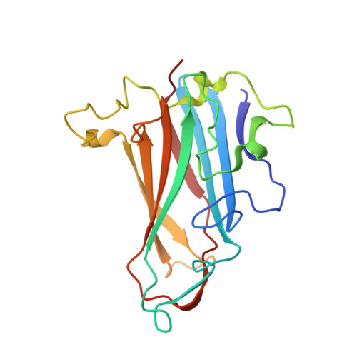
8OFR

Human adenovirus type 25 fiber-knob protein complexed with sialic acid


Experimental Data Snapshot

  • Method: X-RAY DIFFRACTION
  • Resolution: 2.51 Å
  • R-Value Free: 0.239 
  • R-Value Work: 0.221 
  • R-Value Observed: 0.222 

wwPDB Validation   3D Report Full Report


Ligand Structure Quality Assessment 


This is version 1.2 of the entry. See complete history


Literature

Broad sialic acid usage amongst species D human adenovirus.

Mundy, R.M.Baker, A.T.Bates, E.A.Cunliffe, T.G.Teijeira-Crespo, A.Moses, E.Rizkallah, P.J.Parker, A.L.

(2023) Npj Viruses 1: 1-1

  • DOI: https://doi.org/10.1038/s44298-023-00001-5
  • Primary Citation of Related Structures:  
    8OFP, 8OFQ, 8OFR, 8OFS, 8OFT, 8OFU, 8OFV

  • PubMed Abstract: 

    Human adenoviruses (HAdV) are widespread pathogens causing usually mild infections. The Species D (HAdV-D) cause gastrointestinal tract infections and epidemic keratoconjunctivitis (EKC). Despite being significant pathogens, knowledge around HAdV-D mechanism of cell infection is lacking. Sialic acid (SA) usage has been proposed as a cell infection mechanism for EKC causing HAdV-D. Here we highlight an important role for SA engagement by many HAdV-D. We provide apo state crystal structures of 7 previously undetermined HAdV-D fiber-knob proteins, and structures of HAdV-D25, D29, D30 and D53 fiber-knob proteins in complex with SA. Biologically, we demonstrate that removal of cell surface SA reduced infectivity of HAdV-C5 vectors pseudotyped with HAdV-D fiber-knob proteins, whilst engagement of the classical HAdV receptor CAR was variable. Our data indicates variable usage of SA and CAR across HAdV-D. Better defining these interactions will enable improved development of antivirals and engineering of the viruses into refined therapeutic vectors.


  • Organizational Affiliation

    Division of Cancer & Genetics, Cardiff University School of Medicine, Cardiff, CF14 4XN UK.


Macromolecules
Find similar proteins by:  (by identity cutoff)  |  3D Structure
Entity ID: 1
MoleculeChains Sequence LengthOrganismDetailsImage
Fiber
A, B, C, D, E
A, B, C, D, E, F, G, H, I, J, K, L, M, N, O, P, Q, R, S, T, U, V, W, X
186Human adenovirus 25Mutation(s): 0 
Gene Names: L5
UniProt
Find proteins for M0QUM1 (Human adenovirus 25)
Explore M0QUM1 
Go to UniProtKB:  M0QUM1
Entity Groups  
Sequence Clusters30% Identity50% Identity70% Identity90% Identity95% Identity100% Identity
UniProt GroupM0QUM1
Sequence Annotations
Expand
  • Reference Sequence
Small Molecules
Ligands 1 Unique
IDChains Name / Formula / InChI Key2D Diagram3D Interactions
SLB (Subject of Investigation/LOI)
Query on SLB

Download Ideal Coordinates CCD File 
AA [auth C]
BA [auth D]
CA [auth E]
DA [auth F]
EA [auth G]
AA [auth C],
BA [auth D],
CA [auth E],
DA [auth F],
EA [auth G],
FA [auth H],
GA [auth I],
HA [auth J],
IA [auth K],
JA [auth L],
KA [auth M],
LA [auth N],
MA [auth O],
NA [auth P],
OA [auth Q],
PA [auth R],
QA [auth S],
RA [auth T],
SA [auth U],
TA [auth V],
UA [auth W],
VA [auth X],
Y [auth A],
Z [auth B]
N-acetyl-beta-neuraminic acid
C11 H19 N O9
SQVRNKJHWKZAKO-PFQGKNLYSA-N
Experimental Data & Validation

Experimental Data

  • Method: X-RAY DIFFRACTION
  • Resolution: 2.51 Å
  • R-Value Free: 0.239 
  • R-Value Work: 0.221 
  • R-Value Observed: 0.222 
  • Space Group: P 21 21 21
Unit Cell:
Length ( Å )Angle ( ˚ )
a = 127.562α = 90
b = 179.321β = 90
c = 207.897γ = 90
Software Package:
Software NamePurpose
REFMACrefinement
XDSdata reduction
Aimlessdata scaling
PHASERphasing
PDB_EXTRACTdata extraction

Structure Validation

View Full Validation Report


Ligand Structure Quality Assessment 


Entry History & Funding Information

Deposition Data


Funding OrganizationLocationGrant Number
Medical Research Council (MRC, United Kingdom)United KingdomMR/N0137941/1
Wellcome TrustUnited Kingdom517732

Revision History  (Full details and data files)

  • Version 1.0: 2023-09-20
    Type: Initial release
  • Version 1.1: 2023-10-11
    Changes: Database references
  • Version 1.2: 2024-05-08
    Changes: Database references