8PH8

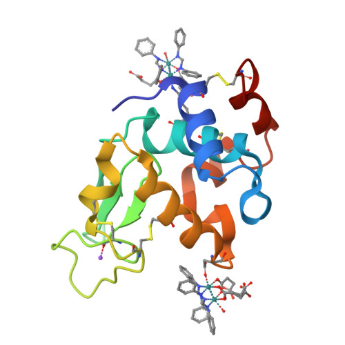
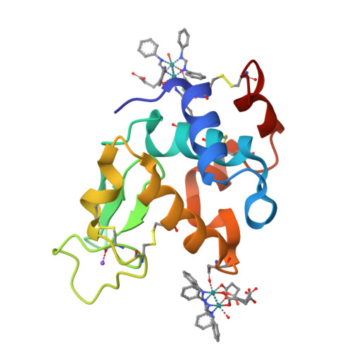
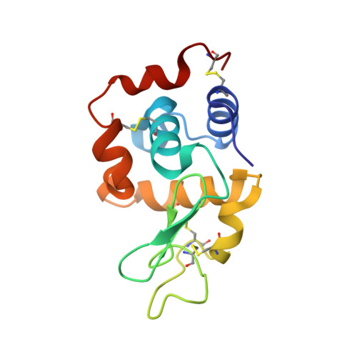
X-ray structure of the adduct formed upon reaction of Lysozyme with [Ru2Cl(DPhF)2(O2CCH3)2] in condition A


Experimental Data Snapshot

  • Method: X-RAY DIFFRACTION
  • Resolution: 1.29 Å
  • R-Value Free: 0.187 
  • R-Value Work: 0.156 

Starting Model: experimental
View more details

wwPDB Validation   3D Report Full Report


Ligand Structure Quality Assessment 

Created with Raphaël 2.3.0Worse 01 BetterLigand structure goodness of fit to experimental dataBest fitted ZJOClick on this verticalbar to view details

This is version 1.2 of the entry. See complete history


Literature

Steric hindrance and charge influence on the cytotoxic activity and protein binding properties of diruthenium complexes.

Teran, A.Ferraro, G.Imbimbo, P.Sanchez-Pelaez, A.E.Monti, D.M.Herrero, S.Merlino, A.

(2023) Int J Biol Macromol 253: 126666-126666

  • DOI: https://doi.org/10.1016/j.ijbiomac.2023.126666
  • Primary Citation of Related Structures:  
    8PH5, 8PH6, 8PH7, 8PH8

  • PubMed Abstract: 

    Paddlewheel diruthenium complexes are being used as metal-based drugs. It has been proposed that their charge and steric properties determine their selectivity towards proteins. Here, we explore these parameters using the first water-soluble diruthenium complex bearing two formamidinate ligands, [Ru 2 Cl(DPhF) 2 (O 2 CCH 3 ) 2 ], and two derivatives, [Ru 2 Cl(DPhF)(O 2 CCH 3 ) 3 ] and K 2 [Ru 2 (DPhF)(CO 3 ) 3 ] (DPhF -  = N,N'-diphenylformamidinate), with one formamidinate. Their protein binding properties have been assessed employing hen egg white lysozyme (HEWL). The results confirm the relationship between the type of interaction (coordinate/non-coordinate bonds) and the charge of diruthenium complexes. The crystallization medium is also a key factor. In all cases, diruthenium species maintain the M-M bond and produce stable adducts. The antiproliferative properties of these diruthenium complexes have been evaluated on an eukaryotic cell-based model. Our data show a correlation between the number of the formamidinate ligands and the anticancer activity of the diruthenium derivatives against human epithelial carcinoma cells. Increased cytotoxicity may be related to increased steric hindrance and Ru 2 5+ core electronic density. However, the effect of increasing the lipophilicity of diruthenium species by introducing a second N,N'-diphenylformamidinate must be also considered. This work illustrates a systematic approach to shed light on the relevant properties of diruthenium compounds to design metal-based metallodrugs and diruthenium metalloenzymes.


  • Organizational Affiliation

    MatMoPol Research Group, Department of Inorganic Chemistry, Faculty of Chemical Sciences, Complutense University of Madrid, Avda. Complutense s/n, 28040 Madrid, Spain.


Macromolecules
Find similar proteins by:  (by identity cutoff)  |  3D Structure
Entity ID: 1
MoleculeChains Sequence LengthOrganismDetailsImage
Lysozyme CA [auth AAA]129Gallus gallusMutation(s): 0 
EC: 3.2.1.17
UniProt
Find proteins for P00698 (Gallus gallus)
Explore P00698 
Go to UniProtKB:  P00698
Entity Groups  
Sequence Clusters30% Identity50% Identity70% Identity90% Identity95% Identity100% Identity
UniProt GroupP00698
Sequence Annotations
Expand
  • Reference Sequence
Small Molecules
Ligands 3 Unique
IDChains Name / Formula / InChI Key2D Diagram3D Interactions
ZJO (Subject of Investigation/LOI)
Query on ZJO

Download Ideal Coordinates CCD File 
D [auth AAA],
E [auth AAA]
1-oxidanyl-2,4,6,8-tetraphenyl-2,4,6,8-tetraza-1$l^{4},5$l^{3}-diruthenabicyclo[3.3.0]octane
C26 H25 N4 O Ru2
PDMORHSIDGQFCT-UHFFFAOYSA-M
SIN
Query on SIN

Download Ideal Coordinates CCD File 
C [auth AAA],
F [auth AAA],
G [auth AAA],
H [auth AAA]
SUCCINIC ACID
C4 H6 O4
KDYFGRWQOYBRFD-UHFFFAOYSA-N
NA
Query on NA

Download Ideal Coordinates CCD File 
B [auth AAA]SODIUM ION
Na
FKNQFGJONOIPTF-UHFFFAOYSA-N
Experimental Data & Validation

Experimental Data

  • Method: X-RAY DIFFRACTION
  • Resolution: 1.29 Å
  • R-Value Free: 0.187 
  • R-Value Work: 0.156 
  • Space Group: P 43 21 2
Unit Cell:
Length ( Å )Angle ( ˚ )
a = 76.89α = 90
b = 76.89β = 90
c = 37.89γ = 90
Software Package:
Software NamePurpose
REFMACrefinement
autoPROCdata reduction
autoPROCdata scaling
PHASERphasing

Structure Validation

View Full Validation Report


Ligand Structure Quality Assessment 

Created with Raphaël 2.3.0Worse 01 BetterLigand structure goodness of fit to experimental dataBest fitted ZJOClick on this verticalbar to view details

Entry History & Funding Information

Deposition Data


Funding OrganizationLocationGrant Number
Not funded--

Revision History  (Full details and data files)

  • Version 1.0: 2023-09-13
    Type: Initial release
  • Version 1.1: 2023-09-20
    Changes: Database references
  • Version 1.2: 2024-10-23
    Changes: Structure summary