8PIZ

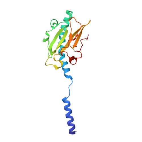
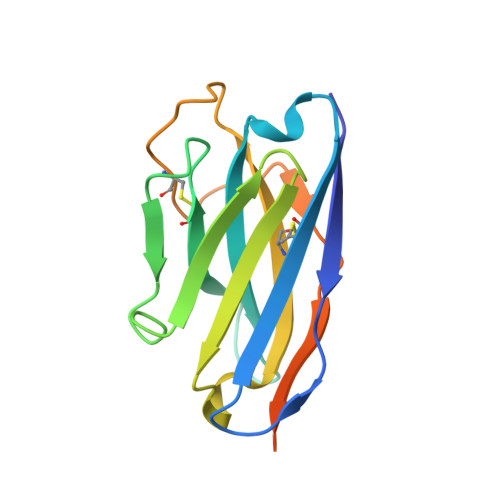
Neisseria meningitidis Type IV pilus SB-DATDH variant bound to the C24 nanobody


Experimental Data Snapshot

  • Method: ELECTRON MICROSCOPY
  • Resolution: 2.75 Å
  • Aggregation State: FILAMENT 
  • Reconstruction Method: HELICAL 

wwPDB Validation   3D Report Full Report


This is version 1.0 of the entry. See complete history


Literature

Cryo-EM structures of type IV pili complexed with nanobodies reveal immune escape mechanisms.

Fernandez-Martinez, D.Kong, Y.Goussard, S.Zavala, A.Gastineau, P.Rey, M.Ayme, G.Chamot-Rooke, J.Lafaye, P.Vos, M.Mechaly, A.Dumenil, G.

(2024) Nat Commun 15: 2414-2414

  • DOI: https://doi.org/10.1038/s41467-024-46677-y
  • Primary Citation of Related Structures:  
    8P2V, 8P36, 8P3B, 8PIJ, 8PIZ, 8PJP

  • PubMed Abstract: 

    Type IV pili (T4P) are prevalent, polymeric surface structures in pathogenic bacteria, making them ideal targets for effective vaccines. However, bacteria have evolved efficient strategies to evade type IV pili-directed antibody responses. Neisseria meningitidis are prototypical type IV pili-expressing Gram-negative bacteria responsible for life threatening sepsis and meningitis. This species has evolved several genetic strategies to modify the surface of its type IV pili, changing pilin subunit amino acid sequence, nature of glycosylation and phosphoforms, but how these modifications affect antibody binding at the structural level is still unknown. Here, to explore this question, we determine cryo-electron microscopy (cryo-EM) structures of pili of different sequence types with sufficiently high resolution to visualize posttranslational modifications. We then generate nanobodies directed against type IV pili which alter pilus function in vitro and in vivo. Cyro-EM in combination with molecular dynamics simulation of the nanobody-pilus complexes reveals how the different types of pili surface modifications alter nanobody binding. Our findings shed light on the impressive complementarity between the different strategies used by bacteria to avoid antibody binding. Importantly, we also show that structural information can be used to make informed modifications in nanobodies as countermeasures to these immune evasion mechanisms.


  • Organizational Affiliation

    Institut Pasteur, Université Paris Cité, INSERM UMR1225, Pathogenesis of Vascular Infections, 75015, Paris, France.


Macromolecules
Find similar proteins by:  (by identity cutoff)  |  3D Structure
Entity ID: 1
MoleculeChains Sequence LengthOrganismDetailsImage
Pilin161Neisseria meningitidis 8013Mutation(s): 0 
UniProt
Find proteins for A0A9K2KM41 (Neisseria meningitidis serogroup C (strain 8013))
Explore A0A9K2KM41 
Go to UniProtKB:  A0A9K2KM41
Entity Groups  
Sequence Clusters30% Identity50% Identity70% Identity90% Identity95% Identity100% Identity
UniProt GroupA0A9K2KM41
Glycosylation
Glycosylation Sites: 1
Sequence Annotations
Expand
  • Reference Sequence
Find similar proteins by:  (by identity cutoff)  |  3D Structure
Entity ID: 2
MoleculeChains Sequence LengthOrganismDetailsImage
C24 nanobody149Vicugna pacosMutation(s): 0 
Entity Groups  
Sequence Clusters30% Identity50% Identity70% Identity90% Identity95% Identity100% Identity
Sequence Annotations
Expand
  • Reference Sequence
Small Molecules
Ligands 2 Unique
IDChains Name / Formula / InChI Key2D Diagram3D Interactions
B6D (Subject of Investigation/LOI)
Query on B6D

Download Ideal Coordinates CCD File 
D [auth A]2,4-bisacetamido-2,4,6-trideoxy-beta-D-glucopyranose
C10 H18 N2 O5
NRXWTRNYICXMBF-DAJIKZSHSA-N
G3P (Subject of Investigation/LOI)
Query on G3P

Download Ideal Coordinates CCD File 
C [auth A]SN-GLYCEROL-3-PHOSPHATE
C3 H9 O6 P
AWUCVROLDVIAJX-GSVOUGTGSA-N
Experimental Data & Validation

Experimental Data

  • Method: ELECTRON MICROSCOPY
  • Resolution: 2.75 Å
  • Aggregation State: FILAMENT 
  • Reconstruction Method: HELICAL 

Structure Validation

View Full Validation Report


Entry History & Funding Information

Deposition Data


Funding OrganizationLocationGrant Number
Agence Nationale de la Recherche (ANR)FranceANR 18 CE11 0022

Revision History  (Full details and data files)

  • Version 1.0: 2024-04-03
    Type: Initial release