8R73 | pdb_00008r73

Polysaccharide lyase BcPL33HA (BcellWH2_04512) Apo form


Experimental Data Snapshot

  • Method: X-RAY DIFFRACTION
  • Resolution: 2.11 Å
  • R-Value Free: 
    0.250 (Depositor), 0.246 (DCC) 
  • R-Value Work: 
    0.189 (Depositor), 0.186 (DCC) 

Starting Model: in silico
View more details

wwPDB Validation   3D Report Full Report


This is version 1.3 of the entry. See complete history


Literature

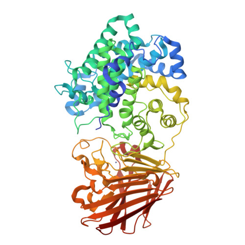
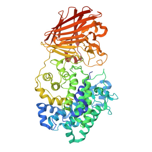
Bacterial polysaccharide lyase family 33: Specificity from an evolutionarily conserved binding tunnel.

Loiodice, M.Drula, E.McIver, Z.Antonyuk, S.Basle, A.Lima, M.Yates, E.A.Byrne, D.P.Coughlan, J.Leech, A.Mesdaghi, S.Rigden, D.J.Drouillard, S.Helbert, W.Henrissat, B.Terrapon, N.Wright, G.S.A.Couturier, M.Cartmell, A.

(2025) Proc Natl Acad Sci U S A 122: e2421623122-e2421623122

  • DOI: https://doi.org/10.1073/pnas.2421623122
  • Primary Citation of Related Structures:  
    8R6Z, 8R70, 8R71, 8R72, 8R73, 8R75

  • PubMed Abstract: 

    Acidic glycans are essential for the biology of multicellular eukaryotes. To utilize them, microbial life including symbionts and pathogens has evolved polysaccharide lyases (PL) that cleave their 1,4 glycosidic linkages via a β-elimination mechanism. PL family 33 (PL33) enzymes have the unusual ability to target a diverse range of glycosaminoglycans (GAGs), as well as the bacterial polymer, gellan gum. In order to gain more detailed insight into PL33 activities we recombinantly expressed 10 PL33 members derived from all major environments and further elucidated the detailed biochemical and biophysical properties of five, showing that their substrate specificity is conferred by variations in tunnel length and topography. The key amino acids involved in catalysis and substrate interactions were identified, and employing a combination of complementary biochemical, structural, and modeling approaches, we show that the tunnel topography is induced by substrate binding to the glycan. Structural and bioinformatic analyses revealed that these features are conserved across several lyase families as well as in mammalian GAG epimerases.


  • Organizational Affiliation

    Université Grenoble Alpes, CNRS, Centre de Recherche sur les Macromolécules Végétales, Grenoble 38000, France.


Macromolecules
Find similar proteins by:  (by identity cutoff)  |  3D Structure
Entity ID: 1
MoleculeChains Sequence LengthOrganismDetailsImage
Heparinase II/III-like protein636Bacteroides cellulosilyticus WH2Mutation(s): 0 
Gene Names: BcellWH2_04512
UniProt
Find proteins for A0A0P0GKX0 (Bacteroides cellulosilyticus)
Explore A0A0P0GKX0 
Go to UniProtKB:  A0A0P0GKX0
Entity Groups  
Sequence Clusters30% Identity50% Identity70% Identity90% Identity95% Identity100% Identity
UniProt GroupA0A0P0GKX0
Sequence Annotations
Expand
  • Reference Sequence
Small Molecules
Ligands 1 Unique
IDChains Name / Formula / InChI Key2D Diagram3D Interactions
ZN
Query on ZN

Download Ideal Coordinates CCD File 
B [auth A]ZINC ION
Zn
PTFCDOFLOPIGGS-UHFFFAOYSA-N
Experimental Data & Validation

Experimental Data

  • Method: X-RAY DIFFRACTION
  • Resolution: 2.11 Å
  • R-Value Free:  0.250 (Depositor), 0.246 (DCC) 
  • R-Value Work:  0.189 (Depositor), 0.186 (DCC) 
Space Group: C 1 2 1
Unit Cell:
Length ( Å )Angle ( ˚ )
a = 103.63α = 90
b = 91.39β = 109.28
c = 84.77γ = 90
Software Package:
Software NamePurpose
REFMACrefinement
xia2data reduction
Aimlessdata scaling
PHASERphasing

Structure Validation

View Full Validation Report


Entry History & Funding Information

Deposition Data

  • Released Date: 2024-12-04 
  • Deposition Author(s): Cartmell, A.

Funding OrganizationLocationGrant Number
Royal SocietyUnited Kingdom212050
Wellcome TrustUnited Kingdom1065163470

Revision History  (Full details and data files)

  • Version 1.0: 2024-12-04
    Type: Initial release
  • Version 1.1: 2025-02-05
    Changes: Structure summary
  • Version 1.2: 2025-02-26
    Changes: Database references
  • Version 1.3: 2025-03-05
    Changes: Database references