8U2M

Structure of P450Blt from Micromonospora sp. MW-13 in Complex with Biarylitide


Experimental Data Snapshot

  • Method: X-RAY DIFFRACTION
  • Resolution: 1.79 Å
  • R-Value Free: 
    0.229 (Depositor), 0.230 (DCC) 
  • R-Value Work: 
    0.199 (Depositor), 0.200 (DCC) 
  • R-Value Observed: 
    0.200 (Depositor) 

Starting Model: experimental
View more details

wwPDB Validation   3D Report Full Report


Ligand Structure Quality Assessment 

Created with Raphaël 2.3.0Worse 01 BetterLigand structure goodness of fit to experimental dataBest fitted HEMClick on this verticalbar to view detailsBest fitted TOEClick on this verticalbar to view details

This is version 1.0 of the entry. See complete history


Literature

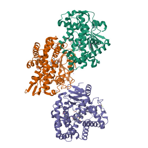
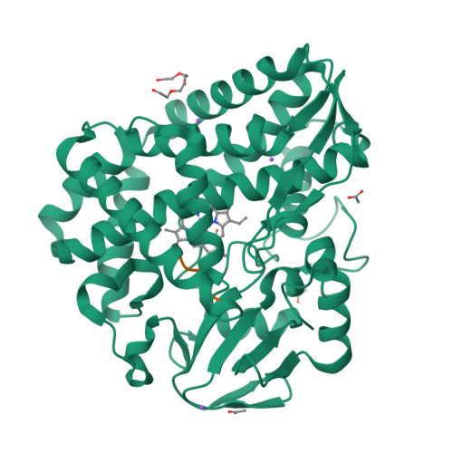
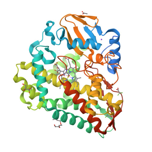
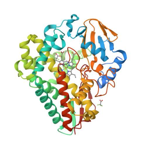
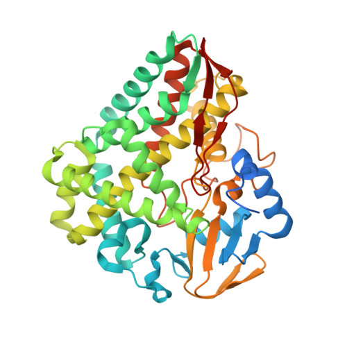
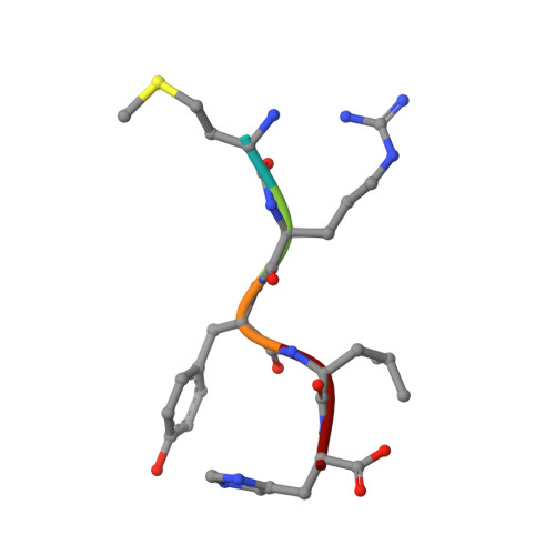
Structural Insights into a Side Chain Cross-Linking Biarylitide P450 from RiPP Biosynthesis

Hansen, M.H.Keto, A.Treisman, M.Sasi, V.M.Coe, L.Zhao, Y.Padva, L.Hess, C.Leichthammer, V.Machell, D.L.Schittenhelm, R.B.Jackson, C.J.Tailhades, J.Crusemann, M.De Voss, J.J.Krenske, E.H.Cryle, M.J.

(2024) ACS Catal : 812-826


Macromolecules
Find similar proteins by:  (by identity cutoff)  |  3D Structure
Entity ID: 1
MoleculeChains Sequence LengthOrganismDetailsImage
Cytochrome P450-SU1
A, B, C
420Micromonospora sp. MW-13Mutation(s): 0 
Gene Names: C5N14_21030
EC: 1.14
UniProt
Find proteins for A0A3E2YLT4 (Micromonospora sp. MW-13)
Explore A0A3E2YLT4 
Go to UniProtKB:  A0A3E2YLT4
Entity Groups  
Sequence Clusters30% Identity50% Identity70% Identity90% Identity95% Identity100% Identity
UniProt GroupA0A3E2YLT4
Sequence Annotations
Expand
  • Reference Sequence

Find similar proteins by:  Sequence   |   3D Structure  

Entity ID: 2
MoleculeChains Sequence LengthOrganismDetailsImage
MET-ARG-TYR-LEU-HIS5synthetic constructMutation(s): 0 
Sequence Annotations
Expand
  • Reference Sequence
Small Molecules
Ligands 7 Unique
IDChains Name / Formula / InChI Key2D Diagram3D Interactions
HEM
Query on HEM

Download Ideal Coordinates CCD File 
J [auth A],
Q [auth B],
V [auth C]
PROTOPORPHYRIN IX CONTAINING FE
C34 H32 Fe N4 O4
KABFMIBPWCXCRK-RGGAHWMASA-L
PG4
Query on PG4

Download Ideal Coordinates CCD File 
G [auth A]TETRAETHYLENE GLYCOL
C8 H18 O5
UWHCKJMYHZGTIT-UHFFFAOYSA-N
TOE
Query on TOE

Download Ideal Coordinates CCD File 
O [auth B]2-[2-(2-METHOXY-ETHOXY)-ETHOXY]-ETHOXYL
C7 H16 O4
JLGLQAWTXXGVEM-UHFFFAOYSA-N
MRY
Query on MRY

Download Ideal Coordinates CCD File 
P [auth B]MESO-ERYTHRITOL
C4 H10 O4
UNXHWFMMPAWVPI-ZXZARUISSA-N
TFA
Query on TFA

Download Ideal Coordinates CCD File 
S [auth B],
T [auth B],
W [auth C],
X [auth C]
trifluoroacetic acid
C2 H F3 O2
DTQVDTLACAAQTR-UHFFFAOYSA-N
ACT
Query on ACT

Download Ideal Coordinates CCD File 
E [auth A]
F [auth A]
H [auth A]
I [auth A]
N [auth B]
ACETATE ION
C2 H3 O2
QTBSBXVTEAMEQO-UHFFFAOYSA-M
NA
Query on NA

Download Ideal Coordinates CCD File 
K [auth A],
L [auth A],
M [auth A],
U [auth B],
Y [auth C]
SODIUM ION
Na
FKNQFGJONOIPTF-UHFFFAOYSA-N
Experimental Data & Validation

Experimental Data

  • Method: X-RAY DIFFRACTION
  • Resolution: 1.79 Å
  • R-Value Free:  0.229 (Depositor), 0.230 (DCC) 
  • R-Value Work:  0.199 (Depositor), 0.200 (DCC) 
  • R-Value Observed: 0.200 (Depositor) 
Space Group: P 1 21 1
Unit Cell:
Length ( Å )Angle ( ˚ )
a = 61.674α = 90
b = 95.607β = 92.71
c = 105.763γ = 90
Software Package:
Software NamePurpose
PHENIXrefinement
Aimlessdata scaling
XDSdata reduction
PHASERphasing

Structure Validation

View Full Validation Report


Ligand Structure Quality Assessment 

Created with Raphaël 2.3.0Worse 01 BetterLigand structure goodness of fit to experimental dataBest fitted HEMClick on this verticalbar to view detailsBest fitted TOEClick on this verticalbar to view details

Entry History & Funding Information

Deposition Data


Funding OrganizationLocationGrant Number
Australian Research Council (ARC)AustraliaDP190101272
Australian Research Council (ARC)AustraliaDP210101752

Revision History  (Full details and data files)

  • Version 1.0: 2024-01-10
    Type: Initial release