8V8E

Room-temperature X-ray structure of SARS-CoV-2 main protease catalytic domain (residues 1-199-6H) in complex with ensitrelvir (ESV)


Experimental Data Snapshot

  • Method: X-RAY DIFFRACTION
  • Resolution: 2.00 Å
  • R-Value Free: 0.193 
  • R-Value Work: 0.149 
  • R-Value Observed: 0.151 

wwPDB Validation   3D Report Full Report


Ligand Structure Quality Assessment 


This is version 1.1 of the entry. See complete history


Literature

Visualizing the Active Site Oxyanion Loop Transition Upon Ensitrelvir Binding and Transient Dimerization of SARS-CoV-2 Main Protease.

Kovalevsky, A.Aniana, A.Coates, L.Ghirlando, R.Nashed, N.T.Louis, J.M.

(2024) J Mol Biol 436: 168616-168616

  • DOI: https://doi.org/10.1016/j.jmb.2024.168616
  • Primary Citation of Related Structures:  
    8V7T, 8V7W, 8V8E, 8V8G

  • PubMed Abstract: 

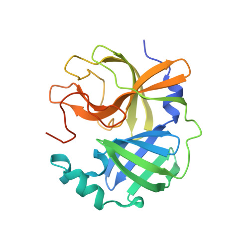
    N-terminal autoprocessing from its polyprotein precursor enables creating the mature-like stable dimer interface of SARS-CoV-2 main protease (MPro), concomitant with the active site oxyanion loop equilibrium transitioning to the active conformation (E*) and onset of catalytic activity. Through mutagenesis of critical interface residues and evaluating noncovalent inhibitor (ensitrelvir, ESV) facilitated dimerization through its weak binding to MPro, we demonstrate that residues extending from Ser1 through Glu14 are critical for dimerization. Combined mutations G11A, E290A and R298A (MPro TM ) restrict dimerization even upon binding of ESV to monomeric MPro TM with an inhibitor dissociation constant of 7.4 ± 1.6 µM. Contrasting the covalent inhibitor NMV or GC373 binding to monomeric MPro, ESV binding enabled capturing the transition of the oxyanion loop conformations in the absence of a reactive warhead and independent of dimerization. Characterization of complexes by room-temperature X-ray crystallography reveals ESV bound to the E* state of monomeric MPro as well as an intermediate approaching the inactive state (E). It appears that the E* to E equilibrium shift occurs initially from G138-F140 residues, leading to the unwinding of the loop and formation of the 3 10 -helix. Finally, we describe a transient dimer structure of the MPro precursor held together through interactions of residues A5-G11 with distinct states of the active sites, E and E*, likely representing an intermediate in the autoprocessing pathway.


  • Organizational Affiliation

    Neutron Scattering Division, Oak Ridge National Laboratory, 1 Bethel Valley Road, Oak Ridge, TN, 37831, USA. Electronic address: kovalevskyay@ornl.gov.


Macromolecules
Find similar proteins by:  (by identity cutoff)  |  3D Structure
Entity ID: 1
MoleculeChains Sequence LengthOrganismDetailsImage
3C-like proteinase nsp5
A, B
207Severe acute respiratory syndrome coronavirus 2Mutation(s): 0 
Gene Names: rep1a-1b
EC: 3.4.22.69
UniProt
Find proteins for P0DTD1 (Severe acute respiratory syndrome coronavirus 2)
Explore P0DTD1 
Go to UniProtKB:  P0DTD1
Entity Groups  
Sequence Clusters30% Identity50% Identity70% Identity90% Identity95% Identity100% Identity
UniProt GroupP0DTD1
Sequence Annotations
Expand
  • Reference Sequence

Find similar proteins by:  Sequence   |   3D Structure  

Entity ID: 2
MoleculeChains Sequence LengthOrganismDetailsImage
peptide9Severe acute respiratory syndrome coronavirus 2Mutation(s): 0 
Sequence Annotations
Expand
  • Reference Sequence
Small Molecules
Ligands 1 Unique
IDChains Name / Formula / InChI Key2D Diagram3D Interactions
7YY (Subject of Investigation/LOI)
Query on 7YY

Download Ideal Coordinates CCD File 
D [auth A],
E [auth B]
6-[(6-chloranyl-2-methyl-indazol-5-yl)amino]-3-[(1-methyl-1,2,4-triazol-3-yl)methyl]-1-[[2,4,5-tris(fluoranyl)phenyl]methyl]-1,3,5-triazine-2,4-dione
C22 H17 Cl F3 N9 O2
QMPBBNUOBOFBFS-UHFFFAOYSA-N
Experimental Data & Validation

Experimental Data

  • Method: X-RAY DIFFRACTION
  • Resolution: 2.00 Å
  • R-Value Free: 0.193 
  • R-Value Work: 0.149 
  • R-Value Observed: 0.151 
  • Space Group: P 1 21 1
Unit Cell:
Length ( Å )Angle ( ˚ )
a = 53.476α = 90
b = 62.641β = 100.23
c = 58.583γ = 90
Software Package:
Software NamePurpose
PHENIXrefinement
CrysalisProdata reduction
CrysalisProdata scaling
PHASERphasing

Structure Validation

View Full Validation Report


Ligand Structure Quality Assessment 


Entry History & Funding Information

Deposition Data


Funding OrganizationLocationGrant Number
Not funded--

Revision History  (Full details and data files)

  • Version 1.0: 2024-05-29
    Type: Initial release
  • Version 1.1: 2024-06-12
    Changes: Database references