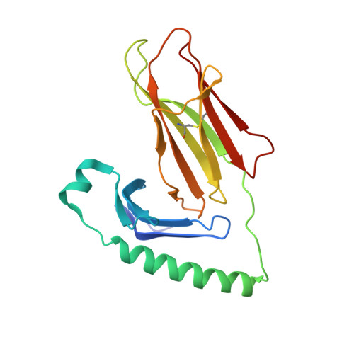
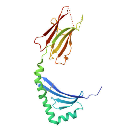
8VDU

Crystal structure of hybrid insulin peptide (InsC8-15-IAPP74-80) bound to HLA-DQ8


Experimental Data Snapshot

  • Method: X-RAY DIFFRACTION
  • Resolution: 3.50 Å
  • R-Value Free: 0.246 
  • R-Value Work: 0.207 
  • R-Value Observed: 0.209 

Starting Model: experimental
View more details

wwPDB Validation   3D Report Full Report


Ligand Structure Quality Assessment 


This is version 1.3 of the entry. See complete history


Literature

A structural basis of T cell cross-reactivity to native and spliced self-antigens presented by HLA-DQ8.

Tran, M.T.Lim, J.J.Loh, T.J.Mannering, S.I.Rossjohn, J.Reid, H.H.

(2024) J Biol Chem 300: 107612-107612

  • DOI: https://doi.org/10.1016/j.jbc.2024.107612
  • Primary Citation of Related Structures:  
    8VCX, 8VCY, 8VD0, 8VD2, 8VDD, 8VDU

  • PubMed Abstract: 

    Type 1 diabetes (T1D) is a T cell-mediated autoimmune disease that has a strong HLA association, where a number of self-epitopes have been implicated in disease pathogenesis. Human pancreatic islet-infiltrating CD4 + T cell clones not only respond to proinsulin C-peptide (PI 40-54; GQVELGGGPGAGSLQ) but also cross-react with a hybrid insulin peptide (HIP; PI 40-47 -IAPP 74-80; GQVELGGG-NAVEVLK) presented by HLA-DQ8. How T cell receptors recognise self-peptide and cross-react to HIPs is unclear. We investigated the cross-reactivity of the CD4 + T cell clones reactive to native PI 40-54 epitope and multiple HIPs fused at the same N-terminus (PI 40-54 ) to the degradation products of two highly expressed pancreatic islet proteins, Neuropeptide Y (NPY 68-74 ) and amyloid polypeptide (IAPP 23-29 and IAPP 74-80 ). We observed that five out of the selected SKW3 T cell lines expressing TCRs isolated from CD4 + T cells of people with T1D responded to multiple HIPs. Despite shared TRAV26-1-TRBV5-1 gene usage in some T cells, these clones cross-reacted to varying degrees with the PI 40-54 and HIP epitopes. Crystal structures of two TRAV26-1 + -TRBV5-1 + T cell receptors (TCRs) in complex with PI 40-54 and HIPs bound to HLA-DQ8 revealed that the two TCRs had distinct mechanisms responsible for their differential recognition of the PI 40-54 and HIP epitopes. Alanine scanning mutagenesis of the PI 40-54 and HIPs determined that the P2, P7 and P8 residues in these epitopes were key determinants of TCR specificity. Accordingly, we provide a molecular basis for cross-reactivity towards native insulin and HIP epitopes presented by HLA-DQ8.


  • Organizational Affiliation

    Infection and Immunity Program & Department of Biochemistry and Molecular Biology, Biomedicine Discovery Institute, Monash University, Clayton, Victoria, Australia 3800.


Macromolecules
Find similar proteins by:  (by identity cutoff)  |  3D Structure
Entity ID: 1
MoleculeChains Sequence LengthOrganismDetailsImage
MHC class II HLA-DQ-alpha chain
A, D, G, J
185Homo sapiensMutation(s): 1 
Gene Names: HLA-DQA1
UniProt
Find proteins for Q30069 (Homo sapiens)
Explore Q30069 
Go to UniProtKB:  Q30069
Entity Groups  
Sequence Clusters30% Identity50% Identity70% Identity90% Identity95% Identity100% Identity
UniProt GroupQ30069
Glycosylation
Glycosylation Sites: 2
Sequence Annotations
Expand
  • Reference Sequence
Find similar proteins by:  (by identity cutoff)  |  3D Structure
Entity ID: 2
MoleculeChains Sequence LengthOrganismDetailsImage
MHC class II HLA-DQ-beta-1
B, E, H, K
192Homo sapiensMutation(s): 0 
Gene Names: HLA-DQB1
UniProt
Find proteins for O19707 (Homo sapiens)
Explore O19707 
Go to UniProtKB:  O19707
Entity Groups  
Sequence Clusters30% Identity50% Identity70% Identity90% Identity95% Identity100% Identity
UniProt GroupO19707
Glycosylation
Glycosylation Sites: 1
Sequence Annotations
Expand
  • Reference Sequence

Find similar proteins by:  Sequence   |   3D Structure  

Entity ID: 3
MoleculeChains Sequence LengthOrganismDetailsImage
Hybrid insulin peptide (HIP; InsC8-15-IAPP74-80)
C, F, I, L
15Homo sapiensMutation(s): 0 
Entity Groups  
Sequence Clusters30% Identity50% Identity70% Identity90% Identity95% Identity100% Identity
Sequence Annotations
Expand
  • Reference Sequence
Oligosaccharides

Help

Entity ID: 4
MoleculeChains Length2D Diagram Glycosylation3D Interactions
beta-D-mannopyranose-(3-4)-2-acetamido-2-deoxy-beta-D-glucopyranose-(1-4)-2-acetamido-2-deoxy-beta-D-glucopyranose
M
3N/AN-Glycosylation
Experimental Data & Validation

Experimental Data

  • Method: X-RAY DIFFRACTION
  • Resolution: 3.50 Å
  • R-Value Free: 0.246 
  • R-Value Work: 0.207 
  • R-Value Observed: 0.209 
  • Space Group: P 1 21 1
Unit Cell:
Length ( Å )Angle ( ˚ )
a = 72.319α = 90
b = 138.116β = 102.498
c = 101.832γ = 90
Software Package:
Software NamePurpose
PHENIXrefinement
autoXDSdata reduction
Cootmodel building
XDSdata scaling
PHASERphasing

Structure Validation

View Full Validation Report


Ligand Structure Quality Assessment 


Entry History & Funding Information

Deposition Data


Funding OrganizationLocationGrant Number
National Health and Medical Research Council (NHMRC, Australia)AustraliaAPP1123586.
National Health and Medical Research Council (NHMRC, Australia)AustraliaAPP1123586

Revision History  (Full details and data files)

  • Version 1.0: 2024-08-07
    Type: Initial release
  • Version 1.1: 2024-08-14
    Changes: Database references
  • Version 1.2: 2024-09-04
    Changes: Database references
  • Version 1.3: 2024-11-20
    Changes: Structure summary