A conserved enzymatic route for azoxy bond formation in natural product biosynthesis
Zang, X., Zhou, J.H., Yiling, D., Jingkun, S., Zhijie, Z., Zhuanglin, S., Guiyun, Z.To be published.
Experimental Data Snapshot
Starting Model: experimental
View more details
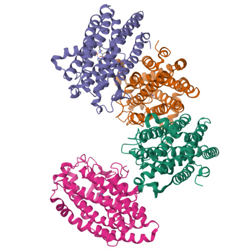
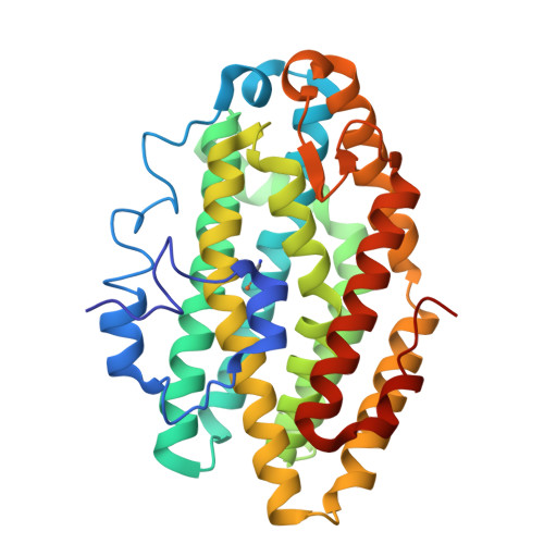
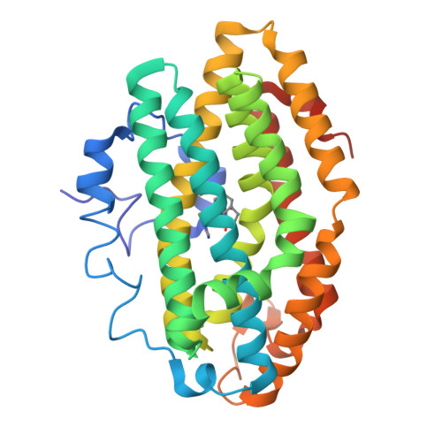
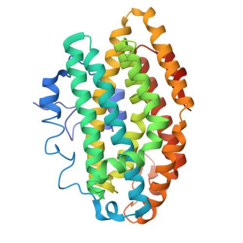
Entity ID: 1 | |||||
|---|---|---|---|---|---|
| Molecule | Chains | Sequence Length | Organism | Details | Image |
| Putative VlmB homolog | 332 | Kitasatospora setae KM-6054 | Mutation(s): 0 Gene Names: vlmB | 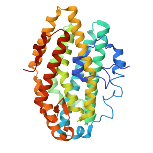 | |
UniProt | |||||
Find proteins for E4N6B3 (Kitasatospora setae (strain ATCC 33774 / DSM 43861 / JCM 3304 / KCC A-0304 / NBRC 14216 / KM-6054)) Explore E4N6B3 Go to UniProtKB: E4N6B3 | |||||
Entity Groups | |||||
| Sequence Clusters | 30% Identity50% Identity70% Identity90% Identity95% Identity100% Identity | ||||
| UniProt Group | E4N6B3 | ||||
Sequence AnnotationsExpand | |||||
| |||||
| Ligands 3 Unique | |||||
|---|---|---|---|---|---|
| ID | Chains | Name / Formula / InChI Key | 2D Diagram | 3D Interactions | |
| XBN (Subject of Investigation/LOI) Query on XBN | L [auth C] | (2~{S})-2-(2-hexylhydrazinyl)-3-oxidanyl-propanoic acid C9 H20 N2 O3 HIBZWEWQIZJYRX-QMMMGPOBSA-N |  | ||
| IMD Query on IMD | E [auth A], H [auth B], K [auth C], O [auth D] | IMIDAZOLE C3 H5 N2 RAXXELZNTBOGNW-UHFFFAOYSA-O |  | ||
| FE Query on FE | F [auth A] G [auth A] I [auth B] J [auth B] M [auth C] | FE (III) ION Fe VTLYFUHAOXGGBS-UHFFFAOYSA-N |  | ||
| Length ( Å ) | Angle ( ˚ ) |
|---|---|
| a = 88.82 | α = 90 |
| b = 95.18 | β = 90 |
| c = 183.25 | γ = 90 |
| Software Name | Purpose |
|---|---|
| PHENIX | refinement |
| XDS | data reduction |
| XDS | data scaling |
| PHASER | phasing |
| Funding Organization | Location | Grant Number |
|---|---|---|
| National Natural Science Foundation of China (NSFC) | China | -- |
RCSB PDB is hosted by

RCSB PDB is a member of the