8YVU

structure of Ige receptor


Experimental Data Snapshot

  • Method: ELECTRON MICROSCOPY
  • Resolution: 3.90 Å
  • Aggregation State: PARTICLE 
  • Reconstruction Method: SINGLE PARTICLE 

wwPDB Validation   3D Report Full Report


This is version 1.0 of the entry. See complete history


Literature

Molecular mechanism of IgE-mediated Fc epsilon RI activation.

Chen, M.Su, Q.Shi, Y.

(2024) Nature 

  • DOI: https://doi.org/10.1038/s41586-024-08229-8
  • Primary Citation of Related Structures:  
    8YVU, 8YWA

  • PubMed Abstract: 

    Allergic diseases, affecting over a quarter of individuals in industrialized countries, have become significant public health concerns 1,2 . The high-affinity Fc receptor for IgE (FcεRI), mainly present on mast cells and basophils, plays a crucial role in allergic diseases 3-5 . Monomeric IgE binding to FcεRI regulates mast cell survival, differentiation, and maturation 6-8 . However, the underlying molecular mechanism remains unclear. Here we demonstrate that, prior to IgE binding, FcεRI mostly exists as a homo-dimer on human mast cell membrane. The structure of human FcεRI confirms the dimeric organization, with each promoter comprising one α subunit, one β subunit, and two γ subunits. The transmembrane helices of the α subunits form a layered arrangement with those of the γ and β subunits. The dimeric interface is mediated by a four-helix bundle of the α and γ subunits at the intracellular juxtamembrane region. Cholesterol-like molecules embedded within the transmembrane domain may stabilize the dimeric assembly. Upon IgE binding, the dimeric FcεRI dissociates into two protomers, each binding to an IgE molecule. Importantly, this process elicits transcriptional activation of Egr1/3 and Ccl2 in rat basophils, which can be attenuated by inhibiting the FcεRI dimer-to-monomer transition. Collectively, our study unveils the mechanism of antigen-independent, IgE-mediated FcεRI activation.


  • Organizational Affiliation

    Research Center for Industries of the Future, Key Laboratory of Structural Biology of Zhejiang Province, School of Life Sciences, Westlake University, Institute of Biology, Westlake Institute for Advanced Study, Xihu District, Hangzhou, Zhejiang Province, China.


Macromolecules
Find similar proteins by:  (by identity cutoff)  |  3D Structure
Entity ID: 1
MoleculeChains Sequence LengthOrganismDetailsImage
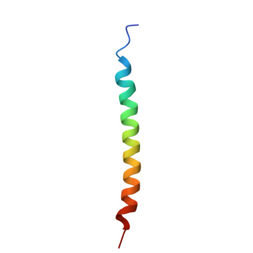
High affinity immunoglobulin epsilon receptor subunit alpha
A, E
37Homo sapiensMutation(s): 0 
Gene Names: FCER1AFCE1A
UniProt & NIH Common Fund Data Resources
Find proteins for P12319 (Homo sapiens)
Explore P12319 
Go to UniProtKB:  P12319
PHAROS:  P12319
GTEx:  ENSG00000179639 
Entity Groups  
Sequence Clusters30% Identity50% Identity70% Identity90% Identity95% Identity100% Identity
UniProt GroupP12319
Sequence Annotations
Expand
  • Reference Sequence
Find similar proteins by:  (by identity cutoff)  |  3D Structure
Entity ID: 2
MoleculeChains Sequence LengthOrganismDetailsImage
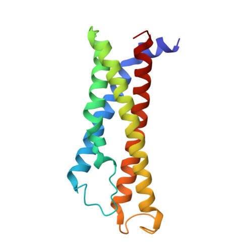
High affinity immunoglobulin epsilon receptor subunit beta
B, F
160Homo sapiensMutation(s): 0 
Gene Names: MS4A2APYFCER1BIGER
UniProt & NIH Common Fund Data Resources
Find proteins for Q01362 (Homo sapiens)
Explore Q01362 
Go to UniProtKB:  Q01362
PHAROS:  Q01362
GTEx:  ENSG00000149534 
Entity Groups  
Sequence Clusters30% Identity50% Identity70% Identity90% Identity95% Identity100% Identity
UniProt GroupQ01362
Sequence Annotations
Expand
  • Reference Sequence
Find similar proteins by:  (by identity cutoff)  |  3D Structure
Entity ID: 3
MoleculeChains Sequence LengthOrganismDetailsImage
High affinity immunoglobulin epsilon receptor subunit gamma
C, D, G, H
39Homo sapiensMutation(s): 0 
Gene Names: FCER1G
UniProt & NIH Common Fund Data Resources
Find proteins for P30273 (Homo sapiens)
Explore P30273 
Go to UniProtKB:  P30273
PHAROS:  P30273
GTEx:  ENSG00000158869 
Entity Groups  
Sequence Clusters30% Identity50% Identity70% Identity90% Identity95% Identity100% Identity
UniProt GroupP30273
Sequence Annotations
Expand
  • Reference Sequence
Experimental Data & Validation

Experimental Data

  • Method: ELECTRON MICROSCOPY
  • Resolution: 3.90 Å
  • Aggregation State: PARTICLE 
  • Reconstruction Method: SINGLE PARTICLE 

Structure Validation

View Full Validation Report


Entry History & Funding Information

Deposition Data


Funding OrganizationLocationGrant Number
National Natural Science Foundation of China (NSFC)China31930059
National Natural Science Foundation of China (NSFC)China81920108015

Revision History  (Full details and data files)

  • Version 1.0: 2024-11-06
    Type: Initial release