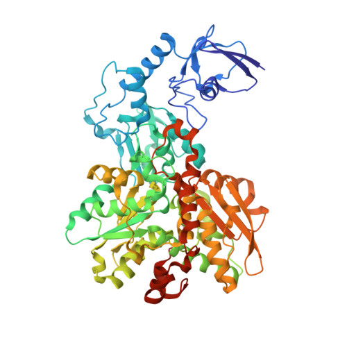
8ALN

CO-bound [FeFe]-hydrogenase I from Clostridium pasteurianum (CpI) at 1.34 Angstrom


Experimental Data Snapshot

  • Method: X-RAY DIFFRACTION
  • Resolution: 1.34 Å
  • R-Value Free: 0.175 
  • R-Value Work: 0.149 
  • R-Value Observed: 0.150 

Starting Model: experimental
View more details

wwPDB Validation   3D Report Full Report


Ligand Structure Quality Assessment 


This is version 2.1 of the entry. See complete history


Literature

Cyanide Binding to [FeFe]-Hydrogenase Stabilizes the Alternative Configuration of the Proton Transfer Pathway.

Duan, J.Hemschemeier, A.Burr, D.J.Stripp, S.T.Hofmann, E.Happe, T.

(2023) Angew Chem Int Ed Engl 62: e202216903-e202216903

  • DOI: https://doi.org/10.1002/anie.202216903
  • Primary Citation of Related Structures:  
    8AIO, 8AJ6, 8ALN, 8AP2

  • PubMed Abstract: 

    Hydrogenases are H 2 converting enzymes that harbor catalytic cofactors in which iron (Fe) ions are coordinated by biologically unusual carbon monoxide (CO) and cyanide (CN - ) ligands. Extrinsic CO and CN - , however, inhibit hydrogenases. The mechanism by which CN - binds to [FeFe]-hydrogenases is not known. Here, we obtained crystal structures of the CN - -treated [FeFe]-hydrogenase CpI from Clostridium pasteurianum. The high resolution of 1.39 Å allowed us to distinguish intrinsic CN - and CO ligands and to show that extrinsic CN - binds to the open coordination site of the cofactor where CO is known to bind. In contrast to other inhibitors, CN - treated crystals show conformational changes of conserved residues within the proton transfer pathway which could allow a direct proton transfer between E279 and S319. This configuration has been proposed to be vital for efficient proton transfer, but has never been observed structurally.


  • Organizational Affiliation

    Department of Plant Biochemistry, Faculty of Biology and Biotechnology, Photobiotechnology, Ruhr University Bochum, Universitätsstrasse 150, 44801, Bochum, Germany.


Macromolecules
Find similar proteins by:  (by identity cutoff)  |  3D Structure
Entity ID: 1
MoleculeChains Sequence LengthOrganismDetailsImage
Iron hydrogenase 1
A, B
584Clostridium pasteurianumMutation(s): 0 
EC: 1.12.7.2
UniProt
Find proteins for P29166 (Clostridium pasteurianum)
Explore P29166 
Go to UniProtKB:  P29166
Entity Groups  
Sequence Clusters30% Identity50% Identity70% Identity90% Identity95% Identity100% Identity
UniProt GroupP29166
Sequence Annotations
Expand
  • Reference Sequence
Small Molecules
Ligands 6 Unique
IDChains Name / Formula / InChI Key2D Diagram3D Interactions
MHX (Subject of Investigation/LOI)
Query on MHX

Download Ideal Coordinates CCD File 
M [auth A],
X [auth B]
Binuclear [FeFe], di(thiomethyl)amine, carbon monoxide, cyanide cluster (-CO form)
C8 H5 Fe2 N3 O4 S2
HQBPXMLEPWPPEN-UHFFFAOYSA-N
SF4 (Subject of Investigation/LOI)
Query on SF4

Download Ideal Coordinates CCD File 
C [auth A]
D [auth A]
E [auth A]
F [auth A]
N [auth B]
C [auth A],
D [auth A],
E [auth A],
F [auth A],
N [auth B],
O [auth B],
P [auth B],
Q [auth B]
IRON/SULFUR CLUSTER
Fe4 S4
LJBDFODJNLIPKO-UHFFFAOYSA-N
FES
Query on FES

Download Ideal Coordinates CCD File 
G [auth A],
R [auth B]
FE2/S2 (INORGANIC) CLUSTER
Fe2 S2
NIXDOXVAJZFRNF-UHFFFAOYSA-N
GOL
Query on GOL

Download Ideal Coordinates CCD File 
I [auth A],
J [auth A],
T [auth B]
GLYCEROL
C3 H8 O3
PEDCQBHIVMGVHV-UHFFFAOYSA-N
CL
Query on CL

Download Ideal Coordinates CCD File 
W [auth B]CHLORIDE ION
Cl
VEXZGXHMUGYJMC-UHFFFAOYSA-M
MG
Query on MG

Download Ideal Coordinates CCD File 
H [auth A]
K [auth A]
L [auth A]
S [auth B]
U [auth B]
H [auth A],
K [auth A],
L [auth A],
S [auth B],
U [auth B],
V [auth B]
MAGNESIUM ION
Mg
JLVVSXFLKOJNIY-UHFFFAOYSA-N
Experimental Data & Validation

Experimental Data

  • Method: X-RAY DIFFRACTION
  • Resolution: 1.34 Å
  • R-Value Free: 0.175 
  • R-Value Work: 0.149 
  • R-Value Observed: 0.150 
  • Space Group: P 1 21 1
Unit Cell:
Length ( Å )Angle ( ˚ )
a = 89.86α = 90
b = 71.85β = 97.47
c = 103.23γ = 90
Software Package:
Software NamePurpose
PHENIXrefinement
XSCALEdata scaling
PDB_EXTRACTdata extraction
XDSdata reduction
PHENIXphasing

Structure Validation

View Full Validation Report


Ligand Structure Quality Assessment 


Entry History & Funding Information

Deposition Data


Funding OrganizationLocationGrant Number
German Research Foundation (DFG)Germany--

Revision History  (Full details and data files)

  • Version 1.0: 2022-12-14
    Type: Initial release
  • Version 1.1: 2023-01-25
    Changes: Database references
  • Version 1.2: 2023-02-15
    Changes: Database references
  • Version 2.0: 2023-03-15
    Changes: Atomic model, Author supporting evidence, Data collection, Derived calculations, Non-polymer description, Structure summary
  • Version 2.1: 2024-02-07
    Changes: Data collection, Refinement description