8R67

tubulin-cryptophycin complex


Experimental Data Snapshot

  • Method: X-RAY DIFFRACTION
  • Resolution: 2.20 Å
  • R-Value Free: 0.229 
  • R-Value Work: 0.194 
  • R-Value Observed: 0.195 

wwPDB Validation   3D Report Full Report


Ligand Structure Quality Assessment 


This is version 1.1 of the entry. See complete history


Literature

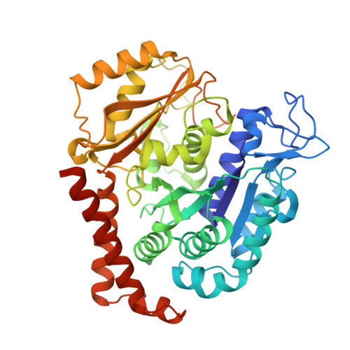
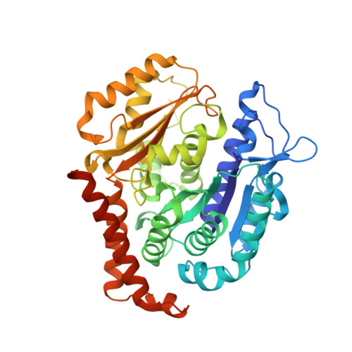
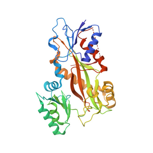
Bridging the maytansine and vinca sites: Cryptophycins target beta-tubulin's T5-loop.

Abel, A.C.Muhlethaler, T.Dessin, C.Schachtsiek, T.Sammet, B.Sharpe, T.Steinmetz, M.O.Sewald, N.Prota, A.E.

(2024) J Biol Chem 300: 107363-107363

  • DOI: https://doi.org/10.1016/j.jbc.2024.107363
  • Primary Citation of Related Structures:  
    8R67

  • PubMed Abstract: 

    Cryptophycins are microtubule-targeting agents (MTAs) that belong to the most potent antimitotic compounds known to date; however, their exact molecular mechanism of action remains unclear. Here, we present the 2.2 Å resolution X-ray crystal structure of a potent cryptophycin derivative bound to the αβ-tubulin heterodimer. The structure addresses conformational issues present in a previous 3.3 Å resolution cryo-electron microscopy structure of cryptophycin-52 bound to the maytansine site of β-tubulin. It further provides atomic details on interactions of cryptophycins, which had not been described previously, including ones that are in line with structure-activity relationship (SAR) studies. Interestingly, we discovered a second cryptophycin-binding site that involves the T5-loop of β-tubulin, a critical secondary structure element involved in the exchange of the guanosine nucleotide and in the formation of longitudinal tubulin contacts in microtubules. Cryptophycins are the first natural ligands found to bind to this new "βT5-loop site" that bridges the maytansine and vinca sites. Our results offer unique avenues to rationally design novel MTAs with the capacity to modulate T5-loop dynamics and to simultaneously engage multiple β-tubulin binding sites.


  • Organizational Affiliation

    Laboratory of Biomolecular Research, Department of Biology and Chemistry, Paul Scherrer Institute, 5232 Villigen PSI, Switzerland; Biozentrum, University of Basel, Spitalstrasse 41, 4056 Basel, Switzerland.


Macromolecules
Find similar proteins by:  (by identity cutoff)  |  3D Structure
Entity ID: 1
MoleculeChains Sequence LengthOrganismDetailsImage
Detyrosinated tubulin alpha-1B chain
A, C
451Bos taurusMutation(s): 0 
UniProt
Find proteins for P81947 (Bos taurus)
Explore P81947 
Go to UniProtKB:  P81947
Entity Groups  
Sequence Clusters30% Identity50% Identity70% Identity90% Identity95% Identity100% Identity
UniProt GroupP81947
Sequence Annotations
Expand
  • Reference Sequence
Find similar proteins by:  (by identity cutoff)  |  3D Structure
Entity ID: 2
MoleculeChains Sequence LengthOrganismDetailsImage
Tubulin beta-2B chain
B, D
445Bos taurusMutation(s): 0 
UniProt
Find proteins for Q6B856 (Bos taurus)
Explore Q6B856 
Go to UniProtKB:  Q6B856
Entity Groups  
Sequence Clusters30% Identity50% Identity70% Identity90% Identity95% Identity100% Identity
UniProt GroupQ6B856
Sequence Annotations
Expand
  • Reference Sequence
Find similar proteins by:  (by identity cutoff)  |  3D Structure
Entity ID: 3
MoleculeChains Sequence LengthOrganismDetailsImage
Stathmin-4143Rattus norvegicusMutation(s): 0 
Gene Names: Stmn4
UniProt
Find proteins for P63043 (Rattus norvegicus)
Explore P63043 
Go to UniProtKB:  P63043
Entity Groups  
Sequence Clusters30% Identity50% Identity70% Identity90% Identity95% Identity100% Identity
UniProt GroupP63043
Sequence Annotations
Expand
  • Reference Sequence
Find similar proteins by:  (by identity cutoff)  |  3D Structure
Entity ID: 4
MoleculeChains Sequence LengthOrganismDetailsImage
Tubulin tyrosine ligase384Gallus gallusMutation(s): 0 
Gene Names: TTL
UniProt
Find proteins for A0A8V0Z8P0 (Gallus gallus)
Explore A0A8V0Z8P0 
Go to UniProtKB:  A0A8V0Z8P0
Entity Groups  
Sequence Clusters30% Identity50% Identity70% Identity90% Identity95% Identity100% Identity
UniProt GroupA0A8V0Z8P0
Sequence Annotations
Expand
  • Reference Sequence
Small Molecules
Ligands 9 Unique
IDChains Name / Formula / InChI Key2D Diagram3D Interactions
Y74 (Subject of Investigation/LOI)
Query on Y74

Download Ideal Coordinates CCD File 
X [auth D],
Y [auth D]
2-[(3~{S},10~{R},13~{E},16~{S})-10-[(3-chloranyl-4-methoxy-phenyl)methyl]-6,6-dimethyl-2,5,9,12-tetrakis(oxidanylidene)-16-[(1~{S})-1-[(2~{R},3~{R})-3-phenyloxiran-2-yl]ethyl]-1,4-dioxa-8,11-diazacyclohexadec-13-en-3-yl]ethanoic acid
C34 H39 Cl N2 O10
GBIUNMWXQNMKGV-AUNCAALYSA-N
GTP
Query on GTP

Download Ideal Coordinates CCD File 
G [auth A],
R [auth C]
GUANOSINE-5'-TRIPHOSPHATE
C10 H16 N5 O14 P3
XKMLYUALXHKNFT-UUOKFMHZSA-N
ACP
Query on ACP

Download Ideal Coordinates CCD File 
CA [auth F]PHOSPHOMETHYLPHOSPHONIC ACID ADENYLATE ESTER
C11 H18 N5 O12 P3
UFZTZBNSLXELAL-IOSLPCCCSA-N
GDP
Query on GDP

Download Ideal Coordinates CCD File 
K [auth B],
V [auth D]
GUANOSINE-5'-DIPHOSPHATE
C10 H15 N5 O11 P2
QGWNDRXFNXRZMB-UUOKFMHZSA-N
MES
Query on MES

Download Ideal Coordinates CCD File 
O [auth B]2-(N-MORPHOLINO)-ETHANESULFONIC ACID
C6 H13 N O4 S
SXGZJKUKBWWHRA-UHFFFAOYSA-N
GOL
Query on GOL

Download Ideal Coordinates CCD File 
P [auth B]GLYCEROL
C3 H8 O3
PEDCQBHIVMGVHV-UHFFFAOYSA-N
IMD
Query on IMD

Download Ideal Coordinates CCD File 
AA [auth E],
M [auth B],
N [auth B],
U [auth C],
Z [auth E]
IMIDAZOLE
C3 H5 N2
RAXXELZNTBOGNW-UHFFFAOYSA-O
CA
Query on CA

Download Ideal Coordinates CCD File 
I [auth A],
J [auth A],
Q [auth B],
T [auth C]
CALCIUM ION
Ca
BHPQYMZQTOCNFJ-UHFFFAOYSA-N
MG
Query on MG

Download Ideal Coordinates CCD File 
BA [auth F],
H [auth A],
L [auth B],
S [auth C],
W [auth D]
MAGNESIUM ION
Mg
JLVVSXFLKOJNIY-UHFFFAOYSA-N
Experimental Data & Validation

Experimental Data

  • Method: X-RAY DIFFRACTION
  • Resolution: 2.20 Å
  • R-Value Free: 0.229 
  • R-Value Work: 0.194 
  • R-Value Observed: 0.195 
  • Space Group: P 21 21 21
Unit Cell:
Length ( Å )Angle ( ˚ )
a = 104.75α = 90
b = 158.66β = 90
c = 180.28γ = 90
Software Package:
Software NamePurpose
PHENIXrefinement
XDSdata reduction
XDSdata scaling
Cootmodel building

Structure Validation

View Full Validation Report


Ligand Structure Quality Assessment 


Entry History & Funding Information

Deposition Data


Funding OrganizationLocationGrant Number
H2020 Marie Curie Actions of the European CommissionEuropean Union860070
Swiss National Science FoundationSwitzerland310030_192566
H2020 Marie Curie Actions of the European CommissionEuropean Union642004

Revision History  (Full details and data files)

  • Version 1.0: 2024-05-22
    Type: Initial release
  • Version 1.1: 2024-06-12
    Changes: Database references