8V30

Smooth Muscle Gamma Actin (ACTG2) Filament Mutant R40C


Experimental Data Snapshot

  • Method: ELECTRON MICROSCOPY
  • Resolution: 2.54 Å
  • Aggregation State: FILAMENT 
  • Reconstruction Method: HELICAL 

wwPDB Validation   3D Report Full Report


This is version 1.1 of the entry. See complete history


Literature

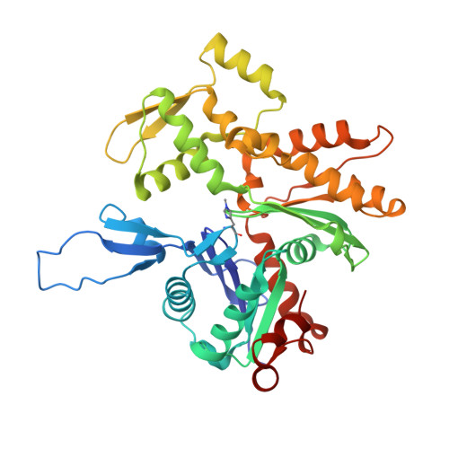
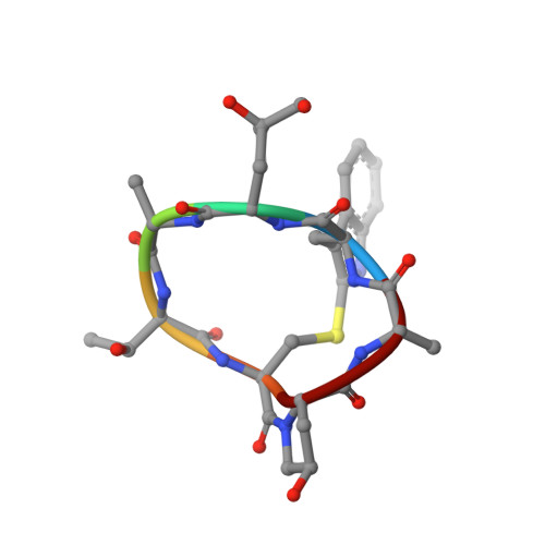
Molecular mechanisms linking missense ACTG2 mutations to visceral myopathy.

Ceron, R.H.Baez-Cruz, F.A.Palmer, N.J.Carman, P.J.Boczkowska, M.Heuckeroth, R.O.Ostap, E.M.Dominguez, R.

(2024) Sci Adv 10: eadn6615-eadn6615

  • DOI: https://doi.org/10.1126/sciadv.adn6615
  • Primary Citation of Related Structures:  
    8V2O, 8V30

  • PubMed Abstract: 

    Visceral myopathy is a life-threatening disease characterized by muscle weakness in the bowel, bladder, and uterus. Mutations in smooth muscle γ-actin (ACTG2) are the most common cause of the disease, but the mechanisms by which the mutations alter muscle function are unknown. Here, we examined four prevalent ACTG2 mutations (R40C, R148C, R178C, and R257C) that cause different disease severity and are spread throughout the actin fold. R178C displayed premature degradation, R148C disrupted interactions with actin-binding proteins, R40C inhibited polymerization, and R257C destabilized filaments. Because these mutations are heterozygous, we also analyzed 50/50 mixtures with wild-type (WT) ACTG2. The WT/R40C mixture impaired filament nucleation by leiomodin 1, and WT/R257C produced filaments that were easily fragmented by smooth muscle myosin. Smooth muscle tropomyosin isoform Tpm1.4 partially rescued the defects of R40C and R257C. Cryo-electron microscopy structures of filaments formed by R40C and R257C revealed disrupted intersubunit contacts. The biochemical and structural properties of the mutants correlate with their genotype-specific disease severity.


  • Organizational Affiliation

    Department of Physiology, Perelman School of Medicine, University of Pennsylvania, Philadelphia, PA 19104, USA.


Macromolecules
Find similar proteins by:  (by identity cutoff)  |  3D Structure
Entity ID: 1
MoleculeChains Sequence LengthOrganismDetailsImage
Actin, gamma-enteric smooth muscle
A, B, C, D, E
376Homo sapiensMutation(s): 1 
Gene Names: ACTG2ACTA3ACTL3ACTSG
EC: 3.6.4
UniProt & NIH Common Fund Data Resources
Find proteins for P63267 (Homo sapiens)
Explore P63267 
Go to UniProtKB:  P63267
PHAROS:  P63267
GTEx:  ENSG00000163017 
Entity Groups  
Sequence Clusters30% Identity50% Identity70% Identity90% Identity95% Identity100% Identity
UniProt GroupP63267
Sequence Annotations
Expand
  • Reference Sequence

Find similar proteins by:  Sequence   |   3D Structure  

Entity ID: 2
MoleculeChains Sequence LengthOrganismDetailsImage
Phalloidin
F, G, H, I, J
7synthetic constructMutation(s): 0 
Sequence Annotations
Expand
  • Reference Sequence
Small Molecules
Ligands 2 Unique
IDChains Name / Formula / InChI Key2D Diagram3D Interactions
ADP
Query on ADP

Download Ideal Coordinates CCD File 
K [auth A],
M [auth B],
P [auth C],
Q [auth D],
S [auth E]
ADENOSINE-5'-DIPHOSPHATE
C10 H15 N5 O10 P2
XTWYTFMLZFPYCI-KQYNXXCUSA-N
MG
Query on MG

Download Ideal Coordinates CCD File 
L [auth A],
N [auth B],
O [auth C],
R [auth D],
T [auth E]
MAGNESIUM ION
Mg
JLVVSXFLKOJNIY-UHFFFAOYSA-N
Modified Residues  2 Unique
IDChains TypeFormula2D DiagramParent
HIC
Query on HIC
A, B, C, D, E
L-PEPTIDE LINKINGC7 H11 N3 O2HIS
HYP
Query on HYP
F, G, H, I, J
L-PEPTIDE LINKINGC5 H9 N O3PRO
Experimental Data & Validation

Experimental Data

  • Method: ELECTRON MICROSCOPY
  • Resolution: 2.54 Å
  • Aggregation State: FILAMENT 
  • Reconstruction Method: HELICAL 
EM Software:
TaskSoftware PackageVersion
RECONSTRUCTIONcryoSPARC4.0
MODEL REFINEMENTPHENIX1.20.1

Structure Validation

View Full Validation Report


Entry History & Funding Information

Deposition Data


Funding OrganizationLocationGrant Number
National Institutes of Health/National Institute of Diabetes and Digestive and Kidney Disease (NIH/NIDDK)United States--

Revision History  (Full details and data files)

  • Version 1.0: 2024-05-01
    Type: Initial release
  • Version 1.1: 2024-06-12
    Changes: Database references