8W1W | pdb_00008w1w

2.03 angstrom resolution crystal structure of as-isolated KatG from Mycobacterium tuberculosis with an MYW-OOH cofactor


Experimental Data Snapshot

  • Method: X-RAY DIFFRACTION
  • Resolution: 2.03 Å
  • R-Value Free: 
    0.223 (Depositor), 0.223 (DCC) 
  • R-Value Work: 
    0.166 (Depositor), 0.166 (DCC) 
  • R-Value Observed: 
    0.166 (Depositor) 

Starting Model: experimental
View more details

wwPDB Validation   3D Report Full Report


Ligand Structure Quality Assessment 

Created with Raphaël 2.3.0Worse 01 BetterLigand structure goodness of fit to experimental dataBest fitted HEMClick on this verticalbar to view details

This is version 1.2 of the entry. See complete history


Literature

Indole N-Linked Hydroperoxyl Adduct of Protein-Derived Cofactor Modulating Catalase-Peroxidase Functions.

Li, J.Duan, R.Traore, E.S.Nguyen, R.C.Davis, I.Griffth, W.P.Goodwin, D.C.Jarzecki, A.A.Liu, A.

(2024) Angew Chem Int Ed Engl 63: e202407018-e202407018

  • DOI: https://doi.org/10.1002/anie.202407018
  • Primary Citation of Related Structures:  
    8CZP, 8U3P, 8W1W, 8W1X, 8W1Y

  • PubMed Abstract: 

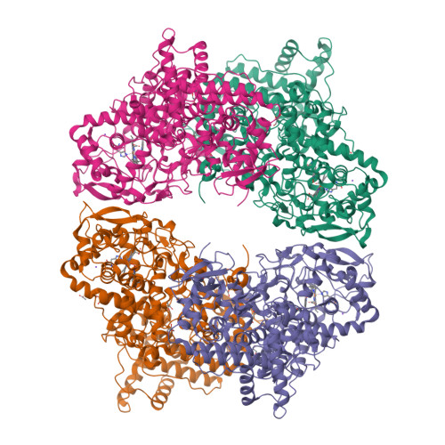
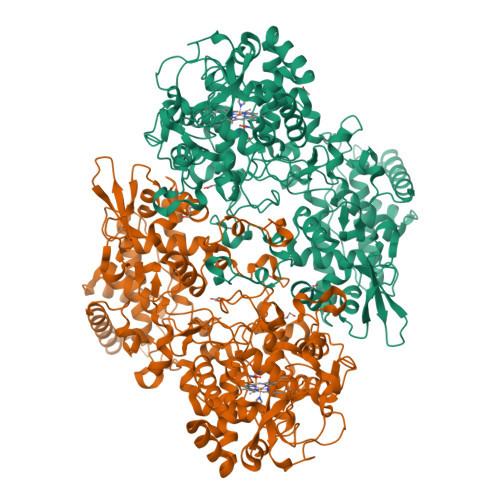
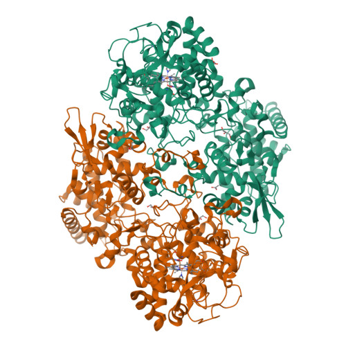
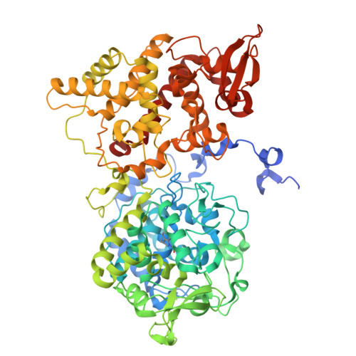
    Bifunctional catalase-peroxidase (KatG) features a posttranslational methionine-tyrosine-tryptophan (MYW) crosslinked cofactor crucial for its catalase function, enabling pathogens to neutralize hydrogen peroxide during infection. We discovered the presence of indole nitrogen-linked hydroperoxyl adduct (MYW-OOH) in Mycobacterium tuberculosis KatG in the solution state under ambient conditions, suggesting its natural occurrence. By isolating predominantly MYW-OOH-containing KatG protein, we investigated the chemical stability and functional impact of MYW-OOH. We discovered that MYW-OOH inhibits catalase activity, presenting a unique temporary lock. Exposure to peroxide or increased temperature removes the hydroperoxyl adduct from the protein cofactor, converting MYW-OOH to MYW and restoring the detoxifying ability of the enzyme against hydrogen peroxide. Thus, the N-linked hydroperoxyl group is releasable. KatG with MYW-OOH represents a catalase dormant, but primed, state of the enzyme. These findings provide insight into chemical strategies targeting the bifunctional enzyme KatG in pathogens, highlighting the role of N-linked hydroperoxyl modifications in enzymatic function.


  • Organizational Affiliation

    University of Texas at San Antonio, Chemistry, UNITED STATES.


Macromolecules
Find similar proteins by:  (by identity cutoff)  |  3D Structure
Entity ID: 1
MoleculeChains Sequence LengthOrganismDetailsImage
Catalase-peroxidase
A, B, C, D
741Mycobacterium tuberculosisMutation(s): 0 
Gene Names: katGERS007661_00994ERS053720_01149
EC: 1.11.1.21
UniProt
Find proteins for P9WIE5 (Mycobacterium tuberculosis (strain ATCC 25618 / H37Rv))
Explore P9WIE5 
Go to UniProtKB:  P9WIE5
Entity Groups  
Sequence Clusters30% Identity50% Identity70% Identity90% Identity95% Identity100% Identity
UniProt GroupP9WIE5
Sequence Annotations
Expand
  • Reference Sequence
Small Molecules
Ligands 4 Unique
IDChains Name / Formula / InChI Key2D Diagram3D Interactions
HEM (Subject of Investigation/LOI)
Query on HEM

Download Ideal Coordinates CCD File 
E [auth A],
J [auth B],
Q [auth C],
V [auth D]
PROTOPORPHYRIN IX CONTAINING FE
C34 H32 Fe N4 O4
KABFMIBPWCXCRK-RGGAHWMASA-L
GOL
Query on GOL

Download Ideal Coordinates CCD File 
G [auth A]
H [auth A]
L [auth B]
M [auth B]
N [auth B]
GLYCEROL
C3 H8 O3
PEDCQBHIVMGVHV-UHFFFAOYSA-N
ACT
Query on ACT

Download Ideal Coordinates CCD File 
I [auth A],
O [auth B],
P [auth B],
Z [auth D]
ACETATE ION
C2 H3 O2
QTBSBXVTEAMEQO-UHFFFAOYSA-M
NA
Query on NA

Download Ideal Coordinates CCD File 
F [auth A],
K [auth B],
R [auth C],
W [auth D]
SODIUM ION
Na
FKNQFGJONOIPTF-UHFFFAOYSA-N
Modified Residues  1 Unique
IDChains TypeFormula2D DiagramParent
TOX
Query on TOX
A, B, C, D
L-PEPTIDE LINKINGC11 H12 N2 O4TRP
Experimental Data & Validation

Experimental Data

  • Method: X-RAY DIFFRACTION
  • Resolution: 2.03 Å
  • R-Value Free:  0.223 (Depositor), 0.223 (DCC) 
  • R-Value Work:  0.166 (Depositor), 0.166 (DCC) 
  • R-Value Observed: 0.166 (Depositor) 
Space Group: P 43 21 2
Unit Cell:
Length ( Å )Angle ( ˚ )
a = 150.674α = 90
b = 150.674β = 90
c = 311.112γ = 90
Software Package:
Software NamePurpose
PHENIXrefinement
DENZOdata reduction
SCALEPACKdata scaling
PDB_EXTRACTdata extraction
PHASERphasing

Structure Validation

View Full Validation Report


Ligand Structure Quality Assessment 

Created with Raphaël 2.3.0Worse 01 BetterLigand structure goodness of fit to experimental dataBest fitted HEMClick on this verticalbar to view details

Entry History & Funding Information

Deposition Data

  • Released Date: 2024-10-23 
  • Deposition Author(s): Liu, A., Li, J.

Funding OrganizationLocationGrant Number
National Institutes of Health/National Institute of General Medical Sciences (NIH/NIGMS)United StatesR01GM108988

Revision History  (Full details and data files)

  • Version 1.0: 2024-10-23
    Type: Initial release
  • Version 1.1: 2024-12-11
    Changes: Database references
  • Version 1.2: 2025-01-08
    Changes: Database references