9BE3

The pre-condensation state of the dimodular NRPS protein LgrA


Experimental Data Snapshot

  • Method: X-RAY DIFFRACTION
  • Resolution: 2.90 Å
  • R-Value Free: 0.258 
  • R-Value Work: 0.216 
  • R-Value Observed: 0.217 

Starting Model: experimental
View more details

wwPDB Validation   3D Report Full Report


Ligand Structure Quality Assessment 

Created with Raphaël 2.3.0Worse 01 BetterLigand structure goodness of fit to experimental dataBest fitted JQGClick on this verticalbar to view detailsBest fitted A1AMOClick on this verticalbar to view details

This is version 1.1 of the entry. See complete history


Literature

Structures and mechanism of condensation in nonribosomal peptide synthesis.

Pistofidis, A.Ma, P.Li, Z.Munro, K.Houk, K.N.Schmeing, T.M.

(2024) Nature 

  • DOI: https://doi.org/10.1038/s41586-024-08417-6
  • Primary Citation of Related Structures:  
    9BE3, 9BE4

  • PubMed Abstract: 

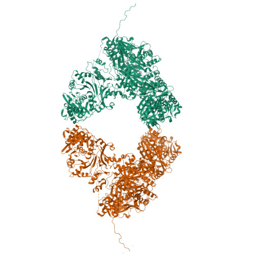
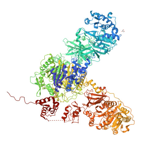
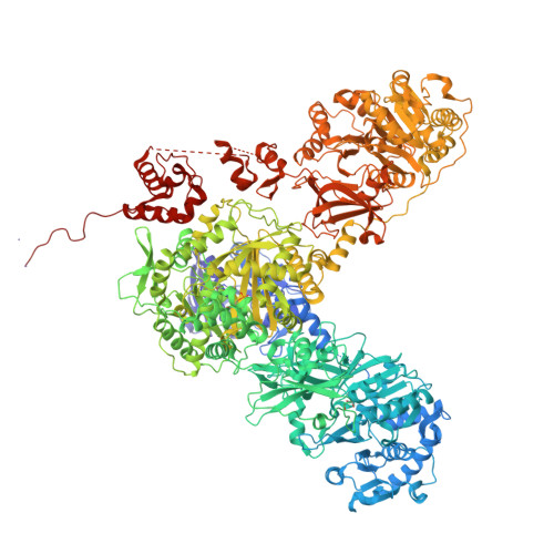
    Nonribosomal peptide synthetases (NRPSs) are mega-enzymes responsible for the biosynthesis of many clinically important natural products, from early modern medicines (penicillin, bacitracin) to current blockbuster drugs (Cubicin, vancomycin) and newly-approved therapeutics (rezafungin) 1,2 . The key chemical step in these biosyntheses is amide bond formation between aminoacyl building blocks, catalyzed by the condensation (C) domain 3 . There has been much debate over the mechanism of this reaction 3-12 . NRPS condensation has been difficult to fully characterize because it is one of many successive reactions in the NRPS synthetic cycle and because the canonical substrates are each attached transiently as thioesters to mobile carrier domains, which are often both contained in the same very flexible protein as the C domain. We have produced a dimodular NRPS protein in two parts, modified each with appropriate non-hydrolysable substrate analogs 13,14 assembled the two parts with protein ligation 15 , and solved structures of the substrate- and product-bound states. The structures show precise orientation of the megaenzyme preparing the nucleophilic attack of its key chemical step, and allow biochemical assays and quantum mechanical simulations to precisely interrogate the reaction. These data suggest that NRPSs C domains use a concerted reaction mechanism, where the active site histidine likely serves not a as general base, but as a crucial stabilizing hydrogen bond acceptor for the developing ammonium.


  • Organizational Affiliation

    Department of Biochemistry and Centre de recherche en biologie structurale, McGill University, Montréal, QC, Canada.


Macromolecules
Find similar proteins by:  (by identity cutoff)  |  3D Structure
Entity ID: 1
MoleculeChains Sequence LengthOrganismDetailsImage
Linear gramicidin synthase subunit A
A, B
1,814Brevibacillus parabrevisMutation(s): 0 
Gene Names: lgrA
UniProt
Find proteins for Q70LM7 (Brevibacillus parabrevis)
Explore Q70LM7 
Go to UniProtKB:  Q70LM7
Entity Groups  
Sequence Clusters30% Identity50% Identity70% Identity90% Identity95% Identity100% Identity
UniProt GroupQ70LM7
Sequence Annotations
Expand
  • Reference Sequence
Small Molecules
Ligands 4 Unique
IDChains Name / Formula / InChI Key2D Diagram3D Interactions
JQG (Subject of Investigation/LOI)
Query on JQG

Download Ideal Coordinates CCD File 
E [auth A],
I [auth B]
(2~{R})-~{N}-[3-[2-[[(2~{S})-2-formamido-3-methyl-butanoyl]amino]ethylamino]-3-oxidanylidene-propyl]-3,3-dimethyl-2-oxidanyl-4-[oxidanyl-bis(oxidanylidene)-$l^{6}-phosphanyl]oxy-butanamide
C17 H32 N4 O9 P
SZJRFDKIBVCSLN-KBPBESRZSA-N
A1AMO (Subject of Investigation/LOI)
Query on A1AMO

Download Ideal Coordinates CCD File 
F [auth A]N-[2-(glycylamino)ethyl]-N~3~-[(2S)-2-hydroxy-3,3-dimethyl-4-(phosphonooxy)butanoyl]-beta-alaninamide
C13 H27 N4 O8 P
ZCWBBGZAVLIBIM-UHFFFAOYSA-N
GOL
Query on GOL

Download Ideal Coordinates CCD File 
C [auth A],
D [auth A],
H [auth B]
GLYCEROL
C3 H8 O3
PEDCQBHIVMGVHV-UHFFFAOYSA-N
MN
Query on MN

Download Ideal Coordinates CCD File 
G [auth A],
J [auth B],
K [auth B],
L [auth B]
MANGANESE (II) ION
Mn
WAEMQWOKJMHJLA-UHFFFAOYSA-N
Experimental Data & Validation

Experimental Data

  • Method: X-RAY DIFFRACTION
  • Resolution: 2.90 Å
  • R-Value Free: 0.258 
  • R-Value Work: 0.216 
  • R-Value Observed: 0.217 
  • Space Group: P 21 21 2
Unit Cell:
Length ( Å )Angle ( ˚ )
a = 184.816α = 90
b = 427.905β = 90
c = 77.318γ = 90
Software Package:
Software NamePurpose
HKL-2000data reduction
HKL-2000data scaling
PHASERphasing
PHENIXrefinement
Cootmodel building

Structure Validation

View Full Validation Report


Ligand Structure Quality Assessment 

Created with Raphaël 2.3.0Worse 01 BetterLigand structure goodness of fit to experimental dataBest fitted JQGClick on this verticalbar to view detailsBest fitted A1AMOClick on this verticalbar to view details

Entry History & Funding Information

Deposition Data


Funding OrganizationLocationGrant Number
Canadian Institutes of Health Research (CIHR)CanadaPJT-178084

Revision History  (Full details and data files)

  • Version 1.0: 2024-11-20
    Type: Initial release
  • Version 1.1: 2024-12-25
    Changes: Database references