9CGL | pdb_00009cgl

Pikromycin Thioesterase Doubly Protected DAP


Experimental Data Snapshot

  • Method: X-RAY DIFFRACTION
  • Resolution: 3.10 Å
  • R-Value Free: 
    0.233 (Depositor), 0.233 (DCC) 
  • R-Value Work: 
    0.181 (Depositor), 0.180 (DCC) 
  • R-Value Observed: 
    0.183 (Depositor) 

Starting Model: experimental
View more details

wwPDB Validation   3D Report Full Report


Ligand Structure Quality Assessment 

Created with Raphaël 2.3.0Worse 01 BetterLigand structure goodness of fit to experimental dataBest fitted A1AWEClick on this verticalbar to view details

This is version 1.1 of the entry. See complete history


Literature

Substrate Trapping in Polyketide Synthase Thioesterase Domains: Structural Basis for Macrolactone Formation.

McCullough, T.M.Choudhary, V.Akey, D.L.Skiba, M.A.Bernard, S.M.Kittendorf, J.D.Schmidt, J.J.Sherman, D.H.Smith, J.L.

(2024) ACS Catal 14: 12551-12563

  • DOI: https://doi.org/10.1021/acscatal.4c03637
  • Primary Citation of Related Structures:  
    9CBD, 9CEL, 9CFJ, 9CGL, 9CGN, 9CGO

  • PubMed Abstract: 

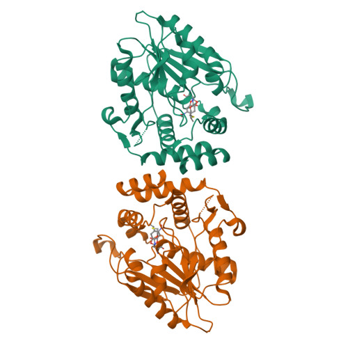
    Emerging antibiotic resistance requires continual improvement in the arsenal of antimicrobial drugs, especially the critical macrolide antibiotics. Formation of the macrolactone scaffold of these polyketide natural products is catalyzed by a modular polyketide synthase (PKS) thioesterase (TE). The TE accepts a linear polyketide substrate from the termina PKS acyl carrier protein to generate an acyl-enzyme adduct that is resolved by attack of a substrate hydroxyl group to form the macrolactone. Our limited mechanistic understanding of TE selectivity for a substrate nucleophile and/or water has hampered development of TEs as biocatalysts that accommodate a variety of natural and non-natural substrates. To understand how TEs direct the substrate nucleophile for macrolactone formation, acyl-enzyme intermediates were trapped as stable amides by substituting the natural serine OH with an amino group. Incorporation of the unnatural amino acid, 1,3-diaminopropionic acid (DAP), was tested with five PKS TEs. DAP-modified TEs (TE DAP ) from the pikromycin and erythromycin pathways were purified and tested with six full-length polyketide intermediates from three pathways. The erythromycin TE had permissive substrate selectivity, whereas the pikromycin TE was selective for its native hexaketide and heptaketide substrates. In a crystal structure of a native substrate trapped in pikromycin TE DAP , the linear heptaketide was curled in the active site with the nucleophilic hydroxyl group positioned 4 Å from the amide-enzyme linkage. The curled heptaketide displayed remarkable shape complementarity with the TE acyl cavity. The strikingly different shapes of acyl cavities in TEs of known structure, including those reported here for juvenimicin, tylosin and fluvirucin biosynthesis, provide insights to facilitate TE engineering and optimization.


  • Organizational Affiliation

    Life Sciences Institute, Mary Sue Coleman Hall, 210 Washtenaw Ave., University of Michigan, Ann Arbor, MI 48109-2216, United States.


Macromolecules
Find similar proteins by:  (by identity cutoff)  |  3D Structure
Entity ID: 1
MoleculeChains Sequence LengthOrganismDetailsImage
Narbonolide/10-deoxymethynolide synthase PikA4, module 6
A, B
290Streptomyces venezuelaeMutation(s): 1 
Gene Names: pikAIV
EC: 2.3.1.239 (PDB Primary Data), 2.3.1.240 (PDB Primary Data)
UniProt
Find proteins for Q9ZGI2 (Streptomyces venezuelae)
Explore Q9ZGI2 
Go to UniProtKB:  Q9ZGI2
Entity Groups  
Sequence Clusters30% Identity50% Identity70% Identity90% Identity95% Identity100% Identity
UniProt GroupQ9ZGI2
Sequence Annotations
Expand
  • Reference Sequence
Small Molecules
Modified Residues  1 Unique
IDChains TypeFormula2D DiagramParent
DPP
Query on DPP
A, B
L-PEPTIDE LINKINGC3 H8 N2 O2ALA
Experimental Data & Validation

Experimental Data

  • Method: X-RAY DIFFRACTION
  • Resolution: 3.10 Å
  • R-Value Free:  0.233 (Depositor), 0.233 (DCC) 
  • R-Value Work:  0.181 (Depositor), 0.180 (DCC) 
  • R-Value Observed: 0.183 (Depositor) 
Space Group: P 21 21 21
Unit Cell:
Length ( Å )Angle ( ˚ )
a = 59.124α = 90
b = 105.858β = 90
c = 114.533γ = 90
Software Package:
Software NamePurpose
PHENIXrefinement
XDSdata reduction
XDSdata scaling
PHENIXphasing

Structure Validation

View Full Validation Report


Ligand Structure Quality Assessment 

Created with Raphaël 2.3.0Worse 01 BetterLigand structure goodness of fit to experimental dataBest fitted A1AWEClick on this verticalbar to view details

Entry History & Funding Information

Deposition Data


Funding OrganizationLocationGrant Number
National Institutes of Health/National Institute of Diabetes and Digestive and Kidney Disease (NIH/NIDDK)United StatesDK042303

Revision History  (Full details and data files)

  • Version 1.0: 2024-09-18
    Type: Initial release
  • Version 1.1: 2025-02-19
    Changes: Database references, Structure summary