Bisected Cap Group Modifications Provides Basis for Improved Pharmacokinetics of HDAC Inhibitors
Olaoye, O., Erdogan, F., Gunning, P.T.To be published.
Experimental Data Snapshot
Starting Model: experimental
View more details
Entity ID: 1 | |||||
|---|---|---|---|---|---|
| Molecule | Chains | Sequence Length | Organism | Details | Image |
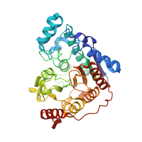
| Hdac6 protein | 357 | Danio rerio | Mutation(s): 0 Gene Names: hdac6 |  | |
UniProt | |||||
Find proteins for A7YT55 (Danio rerio) Explore A7YT55 Go to UniProtKB: A7YT55 | |||||
Entity Groups | |||||
| Sequence Clusters | 30% Identity50% Identity70% Identity90% Identity95% Identity100% Identity | ||||
| UniProt Group | A7YT55 | ||||
Sequence AnnotationsExpand | |||||
| |||||
| Ligands 3 Unique | |||||
|---|---|---|---|---|---|
| ID | Chains | Name / Formula / InChI Key | 2D Diagram | 3D Interactions | |
| A1BH9 (Subject of Investigation/LOI) Query on A1BH9 | C [auth A], F [auth B] | N-hydroxy-4-({[(pyridin-3-yl)methyl](thiophene-3-sulfonyl)amino}methyl)benzamide C18 H17 N3 O4 S2 PMNUORLLCSPXOC-UHFFFAOYSA-N |  | ||
| ZN Query on ZN | D [auth A], G [auth B] | ZINC ION Zn PTFCDOFLOPIGGS-UHFFFAOYSA-N |  | ||
| K Query on K | E [auth A], H [auth B] | POTASSIUM ION K NPYPAHLBTDXSSS-UHFFFAOYSA-N |  | ||
| Length ( Å ) | Angle ( ˚ ) |
|---|---|
| a = 74.79 | α = 90 |
| b = 91.19 | β = 90 |
| c = 96.16 | γ = 90 |
| Software Name | Purpose |
|---|---|
| PHENIX | refinement |
| xia2 | data scaling |
| xia2 | data reduction |
| PHENIX | phasing |
| Funding Organization | Location | Grant Number |
|---|---|---|
| Natural Sciences and Engineering Research Council (NSERC, Canada) | Canada | RGPIN-2014-05767 |
| Canadian Institutes of Health Research (CIHR) | Canada | MOP-130424 |
| Canada Research Chairs | Canada | 950-232042 |
| Ontario Research Fund | Canada | 34876 |
| Canada Foundation for Innovation | Canada | 33536 |