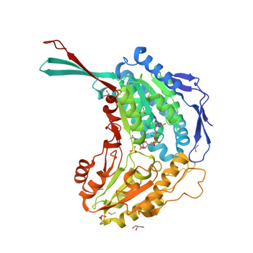
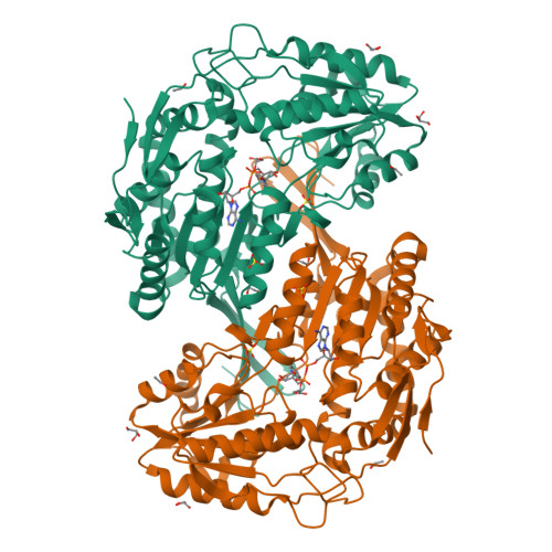
X-ray crystal structure of aldehyde dehydrogenase from Mycobacterium tuberculosis complexed with NAD+.
Moon, J.H., Lyon, A.E., Yu, M., Hung, L.-W., Terwilliger, T., Kim, C.-Y.To be published.
Experimental Data Snapshot
Entity ID: 1 | |||||
|---|---|---|---|---|---|
| Molecule | Chains | Sequence Length | Organism | Details | Image |
| Aldehyde dehydrogenase | 495 | Mycobacterium tuberculosis H37Rv | Mutation(s): 0 Gene Names: Rv0223c, MT0233 EC: 1.2.1 | 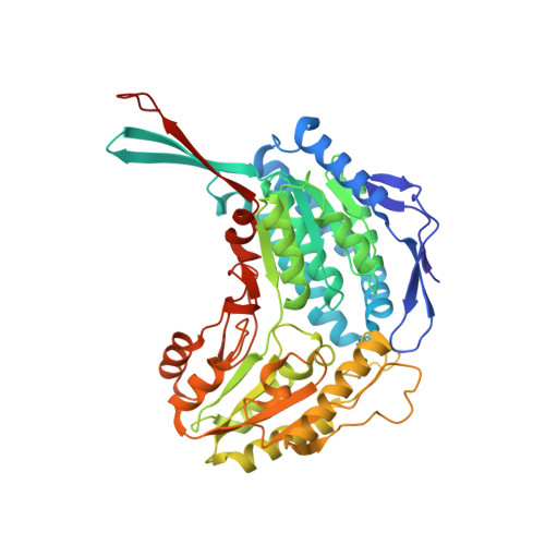 | |
UniProt | |||||
Find proteins for P96405 (Mycobacterium tuberculosis (strain CDC 1551 / Oshkosh)) Explore P96405 Go to UniProtKB: P96405 | |||||
Entity Groups | |||||
| Sequence Clusters | 30% Identity50% Identity70% Identity90% Identity95% Identity100% Identity | ||||
| UniProt Group | P96405 | ||||
Sequence AnnotationsExpand | |||||
| |||||
| Ligands 4 Unique | |||||
|---|---|---|---|---|---|
| ID | Chains | Name / Formula / InChI Key | 2D Diagram | 3D Interactions | |
| NAD Query on NAD | D [auth A] | NICOTINAMIDE-ADENINE-DINUCLEOTIDE C21 H27 N7 O14 P2 BAWFJGJZGIEFAR-NNYOXOHSSA-N |  | ||
| SO4 Query on SO4 | B [auth A], C [auth A] | SULFATE ION O4 S QAOWNCQODCNURD-UHFFFAOYSA-L |  | ||
| GOL Query on GOL | E [auth A], F [auth A], G [auth A], H [auth A] | GLYCEROL C3 H8 O3 PEDCQBHIVMGVHV-UHFFFAOYSA-N |  | ||
| EOH Query on EOH | I [auth A], J [auth A], K [auth A] | ETHANOL C2 H6 O LFQSCWFLJHTTHZ-UHFFFAOYSA-N |  | ||
| Length ( Å ) | Angle ( ˚ ) |
|---|---|
| a = 135.111 | α = 90 |
| b = 135.111 | β = 90 |
| c = 72.543 | γ = 90 |
| Software Name | Purpose |
|---|---|
| PHENIX | refinement |
| ADSC | data collection |
| HKL-2000 | data reduction |
| HKL-2000 | data scaling |
| SOLVE | phasing |
RCSB PDB is hosted by

RCSB PDB is a member of the