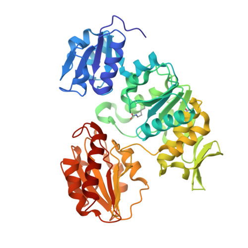
2JFF

Crystal structure of MurD ligase in complex with D-Glu containing sulfonamide inhibitor


Experimental Data Snapshot

  • Method: X-RAY DIFFRACTION
  • Resolution: 1.89 Å
  • R-Value Free: 0.215 
  • R-Value Work: 0.173 
  • R-Value Observed: 0.175 

Starting Model: experimental
View more details

wwPDB Validation   3D Report Full Report


Ligand Structure Quality Assessment 


This is version 1.4 of the entry. See complete history


Literature

Structural and Functional Characterization of Enantiomeric Glutamic Acid Derivatives as Potential Transition State Analogue Inhibitors of Murd Ligase.

Kotnik, M.Humljan, J.Contreras-Martel, C.Oblak, M.Kristan, K.Herve, M.Blanot, D.Urleb, U.Gobec, S.Dessen, A.Solmajer, T.

(2007) J Mol Biol 370: 107

  • DOI: https://doi.org/10.1016/j.jmb.2007.04.048
  • Primary Citation of Related Structures:  
    2JFF, 2JFG, 2JFH

  • PubMed Abstract: 

    Mur ligases play an essential role in the intracellular biosynthesis of bacterial peptidoglycan, the main component of the bacterial cell wall, and represent attractive targets for the design of novel antibacterials. UDP-N-acetylmuramoyl-L-alanine:D-glutamate ligase (MurD) catalyses the addition of D-glutamic acid to the cytoplasmic intermediate UDP-N-acetylmuramoyl-L-alanine (UMA) and is the second in the series of Mur ligases. MurD ligase is highly stereospecific for its substrate, D-glutamic acid (D-Glu). Here, we report the high resolution crystal structures of MurD in complexes with two novel inhibitors designed to mimic the transition state of the reaction, which contain either the D-Glu or the L-Glu moiety. The binding modes of N-sulfonyl-D-Glu and N-sulfonyl-L-Glu derivatives were also characterised kinetically. The results of this study represent an excellent starting point for further development of novel inhibitors of this enzyme.


  • Organizational Affiliation

    Lek Pharmaceuticals d. d., Drug Discovery, Verovskova 57, 1526 Ljubljana, Slovenia. miha.kotnik@sandoz.com


Macromolecules
Find similar proteins by:  (by identity cutoff)  |  3D Structure
Entity ID: 1
MoleculeChains Sequence LengthOrganismDetailsImage
UDP-N-ACETYLMURAMOYLALANINE--D-GLUTAMATE LIGASE445Escherichia coliMutation(s): 0 
EC: 6.3.2.9
UniProt
Find proteins for P14900 (Escherichia coli (strain K12))
Explore P14900 
Go to UniProtKB:  P14900
Entity Groups  
Sequence Clusters30% Identity50% Identity70% Identity90% Identity95% Identity100% Identity
UniProt GroupP14900
Sequence Annotations
Expand
  • Reference Sequence
Small Molecules
Modified Residues  1 Unique
IDChains TypeFormula2D DiagramParent
KCX
Query on KCX
A
L-PEPTIDE LINKINGC7 H14 N2 O4LYS
Binding Affinity Annotations 
IDSourceBinding Affinity
LK2 BindingDB:  2JFF Kd: min: 1.20e+5, max: 2.10e+5 (nM) from 2 assay(s)
IC50: 2.80e+5 (nM) from 1 assay(s)
Experimental Data & Validation

Experimental Data

  • Method: X-RAY DIFFRACTION
  • Resolution: 1.89 Å
  • R-Value Free: 0.215 
  • R-Value Work: 0.173 
  • R-Value Observed: 0.175 
  • Space Group: P 41
Unit Cell:
Length ( Å )Angle ( ˚ )
a = 65.288α = 90
b = 65.288β = 90
c = 135.06γ = 90
Software Package:
Software NamePurpose
REFMACrefinement
XDSdata reduction
XSCALEdata scaling
PHASERphasing

Structure Validation

View Full Validation Report


Ligand Structure Quality Assessment 


Entry History 

Deposition Data

Revision History  (Full details and data files)

  • Version 1.0: 2007-05-15
    Type: Initial release
  • Version 1.1: 2011-05-08
    Changes: Version format compliance
  • Version 1.2: 2011-07-13
    Changes: Version format compliance
  • Version 1.3: 2018-01-31
    Changes: Source and taxonomy
  • Version 1.4: 2023-12-13
    Changes: Data collection, Database references, Derived calculations, Other, Refinement description