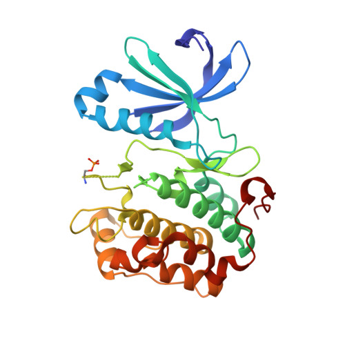
2PE1

CRYSTAL STRUCTURE OF HUMAN PHOSPHOINOSITIDE-DEPENDENT PROTEIN KINASE 1 (PDK1) {2-Oxo-3-[1-(1H-pyrrol-2-yl)-eth-(Z)-ylidene]-2,3-dihydro-1H-indol-5-yl}-urea {BX-517} COMPLEX


Experimental Data Snapshot

  • Method: X-RAY DIFFRACTION
  • Resolution: 2.14 Å
  • R-Value Free: 0.282 
  • R-Value Work: 0.222 
  • R-Value Observed: 0.224 

wwPDB Validation   3D Report Full Report


Ligand Structure Quality Assessment 


This is version 1.3 of the entry. See complete history


Literature

Indolinone based phosphoinositide-dependent kinase-1 (PDK1) inhibitors. Part 1: Design, synthesis and biological activity.

Islam, I.Bryant, J.Chou, Y.L.Kochanny, M.J.Lee, W.Phillips, G.B.Yu, H.Adler, M.Whitlow, M.Ho, E.Lentz, D.Polokoff, M.A.Subramanyam, B.Wu, J.M.Zhu, D.Feldman, R.I.Arnaiz, D.O.

(2007) Bioorg Med Chem Lett 17: 3814-3818

  • DOI: https://doi.org/10.1016/j.bmcl.2007.04.071
  • Primary Citation of Related Structures:  
    2PE0, 2PE1

  • PubMed Abstract: 

    HTS screening identified 1 with micromolar inhibitory activity against PDK1. Optimization of 1 afforded 4i (BX-517) which has single-digit nanomolar activity against PDK1 and excellent selectivity against PKA.


  • Organizational Affiliation

    Berlex Biosciences, 2600 Hilltop Dr. Richmond, CA 94804, USA.


Macromolecules
Find similar proteins by:  (by identity cutoff)  |  3D Structure
Entity ID: 1
MoleculeChains Sequence LengthOrganismDetailsImage
3-phosphoinositide-dependent protein kinase 1286Homo sapiensMutation(s): 1 
Gene Names: PDPK1PDK1
EC: 2.7.11.1
UniProt & NIH Common Fund Data Resources
Find proteins for O15530 (Homo sapiens)
Explore O15530 
Go to UniProtKB:  O15530
PHAROS:  O15530
GTEx:  ENSG00000140992 
Entity Groups  
Sequence Clusters30% Identity50% Identity70% Identity90% Identity95% Identity100% Identity
UniProt GroupO15530
Sequence Annotations
Expand
  • Reference Sequence
Small Molecules
Ligands 3 Unique
IDChains Name / Formula / InChI Key2D Diagram3D Interactions
517
Query on 517

Download Ideal Coordinates CCD File 
D [auth A]1-{2-OXO-3-[(1R)-1-(1H-PYRROL-2-YL)ETHYL]-2H-INDOL-5-YL}UREA
C15 H14 N4 O2
KJDBLWKTHMHALD-QMMMGPOBSA-N
SO4
Query on SO4

Download Ideal Coordinates CCD File 
B [auth A],
C [auth A]
SULFATE ION
O4 S
QAOWNCQODCNURD-UHFFFAOYSA-L
GOL
Query on GOL

Download Ideal Coordinates CCD File 
E [auth A],
F [auth A],
G [auth A],
H [auth A]
GLYCEROL
C3 H8 O3
PEDCQBHIVMGVHV-UHFFFAOYSA-N
Modified Residues  1 Unique
IDChains TypeFormula2D DiagramParent
SEP
Query on SEP
A
L-PEPTIDE LINKINGC3 H8 N O6 PSER
Binding Affinity Annotations 
IDSourceBinding Affinity
517 PDBBind:  2PE1 IC50: 5 (nM) from 1 assay(s)
Experimental Data & Validation

Experimental Data

  • Method: X-RAY DIFFRACTION
  • Resolution: 2.14 Å
  • R-Value Free: 0.282 
  • R-Value Work: 0.222 
  • R-Value Observed: 0.224 
  • Space Group: P 32 2 1
Unit Cell:
Length ( Å )Angle ( ˚ )
a = 125.234α = 90
b = 125.234β = 90
c = 47.696γ = 120
Software Package:
Software NamePurpose
CNXrefinement
XTALVIEWrefinement
X-GENdata reduction
X-GENdata scaling
CNXphasing

Structure Validation

View Full Validation Report


Ligand Structure Quality Assessment 


Entry History 

Deposition Data

Revision History  (Full details and data files)

  • Version 1.0: 2007-06-12
    Type: Initial release
  • Version 1.1: 2008-05-01
    Changes: Version format compliance
  • Version 1.2: 2011-07-13
    Changes: Non-polymer description, Version format compliance
  • Version 1.3: 2024-11-06
    Changes: Data collection, Database references, Derived calculations, Structure summary