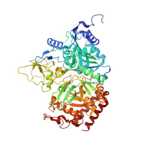
3DTB

The structure of rat cytosolic PEPCK in complex with phosphoglycolate and GDP


Experimental Data Snapshot

  • Method: X-RAY DIFFRACTION
  • Resolution: 1.30 Å
  • R-Value Free: 0.193 
  • R-Value Work: 0.168 
  • R-Value Observed: 0.169 

wwPDB Validation   3D Report Full Report


Ligand Structure Quality Assessment 


This is version 1.3 of the entry. See complete history


Literature

Enzymes with lid-gated active sites must operate by an induced fit mechanism instead of conformational selection.

Sullivan, S.M.Holyoak, T.

(2008) Proc Natl Acad Sci U S A 105: 13829-13834

  • DOI: https://doi.org/10.1073/pnas.0805364105
  • Primary Citation of Related Structures:  
    3DT2, 3DT4, 3DT7, 3DTB

  • PubMed Abstract: 

    The induced fit and conformational selection/population shift models are two extreme cases of a continuum aimed at understanding the mechanism by which the final key-lock or active enzyme conformation is achieved upon formation of the correctly ligated enzyme. Structures of complexes representing the Michaelis and enolate intermediate complexes of the reaction catalyzed by phosphoenolpyruvate carboxykinase provide direct structural evidence for the encounter complex that is intrinsic to the induced fit model and not required by the conformational selection model. In addition, the structural data demonstrate that the conformational selection model is not sufficient to explain the correlation between dynamics and catalysis in phosphoenolpyruvate carboxykinase and other enzymes in which the transition between the uninduced and the induced conformations occludes the active site from the solvent. The structural data are consistent with a model in that the energy input from substrate association results in changes in the free energy landscape for the protein, allowing for structural transitions along an induced fit pathway.


  • Organizational Affiliation

    Department of Biochemistry and Molecular Biology, University of Kansas Medical Center, Kansas City, KS 66160, USA.


Macromolecules
Find similar proteins by:  (by identity cutoff)  |  3D Structure
Entity ID: 1
MoleculeChains Sequence LengthOrganismDetailsImage
Phosphoenolpyruvate carboxykinase, cytosolic [GTP]
A, B
624Rattus norvegicusMutation(s): 0 
Gene Names: Pck1
EC: 4.1.1.32 (PDB Primary Data), 2.7.11 (UniProt)
UniProt
Find proteins for P07379 (Rattus norvegicus)
Explore P07379 
Go to UniProtKB:  P07379
Entity Groups  
Sequence Clusters30% Identity50% Identity70% Identity90% Identity95% Identity100% Identity
UniProt GroupP07379
Sequence Annotations
Expand
  • Reference Sequence
Small Molecules
Ligands 7 Unique
IDChains Name / Formula / InChI Key2D Diagram3D Interactions
GDP
Query on GDP

Download Ideal Coordinates CCD File 
H [auth A],
P [auth B]
GUANOSINE-5'-DIPHOSPHATE
C10 H15 N5 O11 P2
QGWNDRXFNXRZMB-UUOKFMHZSA-N
EPE
Query on EPE

Download Ideal Coordinates CCD File 
K [auth A]4-(2-HYDROXYETHYL)-1-PIPERAZINE ETHANESULFONIC ACID
C8 H18 N2 O4 S
JKMHFZQWWAIEOD-UHFFFAOYSA-N
1PE
Query on 1PE

Download Ideal Coordinates CCD File 
I [auth A],
J [auth A]
PENTAETHYLENE GLYCOL
C10 H22 O6
JLFNLZLINWHATN-UHFFFAOYSA-N
PGA
Query on PGA

Download Ideal Coordinates CCD File 
C [auth A],
L [auth B]
2-PHOSPHOGLYCOLIC ACID
C2 H5 O6 P
ASCFNMCAHFUBCO-UHFFFAOYSA-N
BCT
Query on BCT

Download Ideal Coordinates CCD File 
G [auth A]BICARBONATE ION
C H O3
BVKZGUZCCUSVTD-UHFFFAOYSA-M
MN
Query on MN

Download Ideal Coordinates CCD File 
D [auth A],
E [auth A],
M [auth B],
N [auth B]
MANGANESE (II) ION
Mn
WAEMQWOKJMHJLA-UHFFFAOYSA-N
NA
Query on NA

Download Ideal Coordinates CCD File 
F [auth A],
O [auth B]
SODIUM ION
Na
FKNQFGJONOIPTF-UHFFFAOYSA-N
Experimental Data & Validation

Experimental Data

  • Method: X-RAY DIFFRACTION
  • Resolution: 1.30 Å
  • R-Value Free: 0.193 
  • R-Value Work: 0.168 
  • R-Value Observed: 0.169 
  • Space Group: P 1 21 1
Unit Cell:
Length ( Å )Angle ( ˚ )
a = 62.154α = 90
b = 119.683β = 107.05
c = 86.849γ = 90
Software Package:
Software NamePurpose
DENZOdata reduction
SCALEPACKdata scaling
REFMACrefinement
PDB_EXTRACTdata extraction
Blu-Icedata collection
HKL-2000data reduction
HKL-2000data scaling

Structure Validation

View Full Validation Report


Ligand Structure Quality Assessment 


Entry History 

Deposition Data

Revision History  (Full details and data files)

  • Version 1.0: 2008-08-26
    Type: Initial release
  • Version 1.1: 2011-07-13
    Changes: Advisory, Version format compliance
  • Version 1.2: 2017-10-25
    Changes: Refinement description
  • Version 1.3: 2024-02-21
    Changes: Data collection, Database references, Derived calculations