3QS6

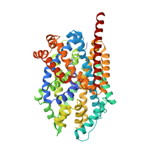
Crystal structure of LeuT mutant F259V,I359Q bound to sodium and L-tryptophan


Experimental Data Snapshot

  • Method: X-RAY DIFFRACTION
  • Resolution: 2.80 Å
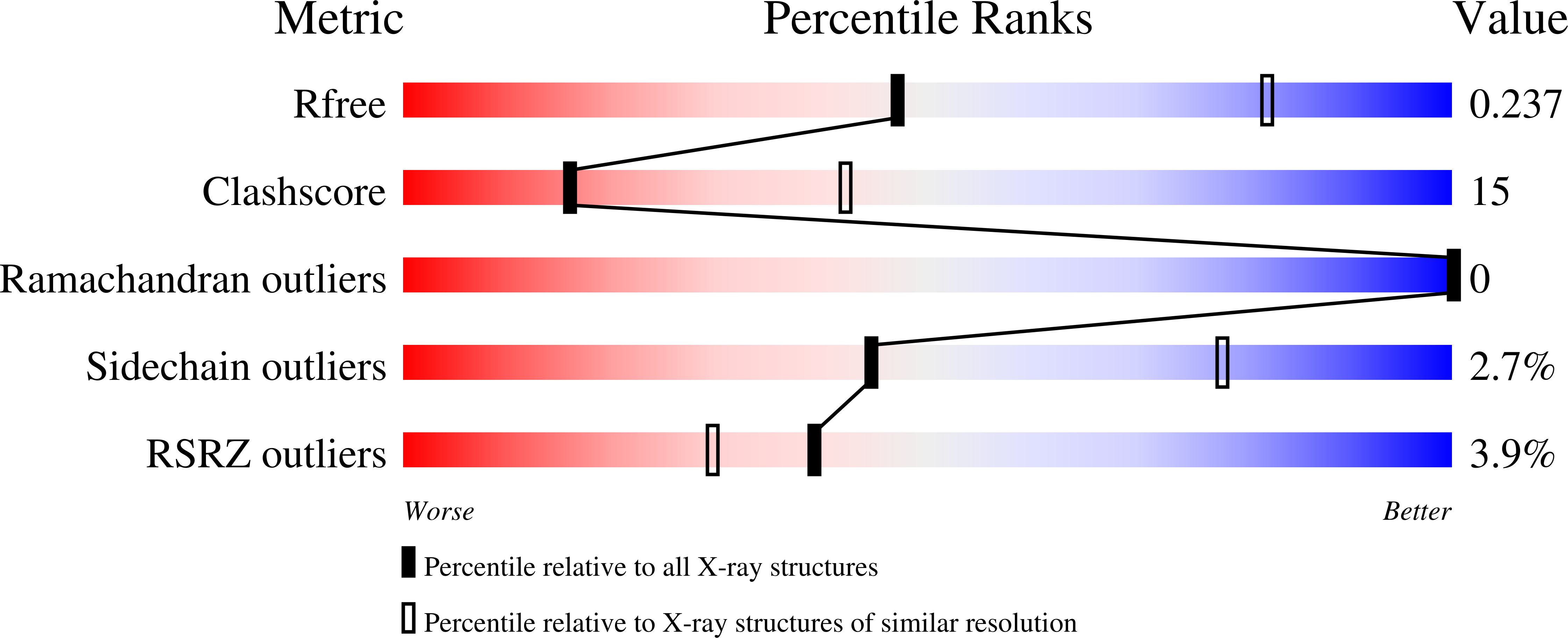
  • R-Value Free: 0.237 
  • R-Value Work: 0.200 
  • R-Value Observed: 0.202 

Starting Model: experimental
View more details

wwPDB Validation   3D Report Full Report


Ligand Structure Quality Assessment 


This is version 1.3 of the entry. See complete history


Literature

Insights into transport mechanism from LeuT engineered to transport tryptophan.

Piscitelli, C.L.Gouaux, E.

(2012) EMBO J 31: 228-235

  • DOI: https://doi.org/10.1038/emboj.2011.353
  • Primary Citation of Related Structures:  
    3QS4, 3QS5, 3QS6

  • PubMed Abstract: 

    LeuT is a bacterial homologue of the neurotransmitter:sodium symporter (NSS) family and, being the only NSS member to have been structurally characterized by X-ray crystallography, is a model protein for studying transporter structure and mechanism. Transport activity in LeuT was hypothesized to require structural transitions between open-to-out and occluded conformations dependent upon protein:ligand binding complementarity. Here, using crystallographic and functional analysis, we show that binding site modification produces changes in both structure and activity that are consistent with complementarity-dependent structural transitions to the occluded state. The mutation I359Q converts the activity of tryptophan from inhibitor to transportable substrate. This mutation changes the local environment of the binding site, inducing the bound tryptophan to adopt a different conformer than in the wild-type complex. Instead of trapping the transporter open, tryptophan binding now allows the formation of an occluded state. Thus, transport activity is correlated to the ability of the ligand to promote the structural transition to the occluded state, a step in the transport cycle that is dependent on protein:ligand complementarity in the central binding site.


  • Organizational Affiliation

    Department of Biochemistry and Molecular Biology, Oregon Health and Science University, Portland, OR 97239, USA.


Macromolecules
Find similar proteins by:  (by identity cutoff)  |  3D Structure
Entity ID: 1
MoleculeChains Sequence LengthOrganismDetailsImage
Na(+):neurotransmitter symporter (Snf family)519Aquifex aeolicusMutation(s): 2 
Gene Names: aq_2077leutsnf
Membrane Entity: Yes 
UniProt
Find proteins for O67854 (Aquifex aeolicus (strain VF5))
Explore O67854 
Go to UniProtKB:  O67854
Entity Groups  
Sequence Clusters30% Identity50% Identity70% Identity90% Identity95% Identity100% Identity
UniProt GroupO67854
Sequence Annotations
Expand
  • Reference Sequence
Small Molecules
Binding Affinity Annotations 
IDSourceBinding Affinity
TRP PDBBind:  3QS6 Kd: 1.28e+4 (nM) from 1 assay(s)
Experimental Data & Validation

Experimental Data

  • Method: X-RAY DIFFRACTION
  • Resolution: 2.80 Å
  • R-Value Free: 0.237 
  • R-Value Work: 0.200 
  • R-Value Observed: 0.202 
  • Space Group: C 1 2 1
Unit Cell:
Length ( Å )Angle ( ˚ )
a = 91.793α = 90
b = 87.127β = 94.73
c = 82.169γ = 90
Software Package:
Software NamePurpose
ADSCdata collection
MOLREPphasing
PHENIXrefinement
HKL-2000data reduction
HKL-2000data scaling

Structure Validation

View Full Validation Report


Ligand Structure Quality Assessment 


Entry History 

Deposition Data

Revision History  (Full details and data files)

  • Version 1.0: 2011-10-12
    Type: Initial release
  • Version 1.1: 2012-02-29
    Changes: Database references
  • Version 1.2: 2020-07-29
    Type: Remediation
    Reason: Carbohydrate remediation
    Changes: Data collection, Database references, Derived calculations, Structure summary
  • Version 1.3: 2023-09-13
    Changes: Data collection, Database references, Refinement description, Structure summary