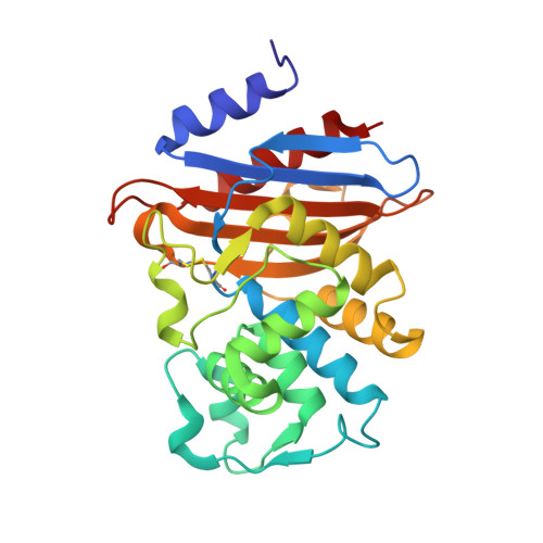
3RXW

KPC-2 carbapenemase in complex with PSR3-226


Experimental Data Snapshot

  • Method: X-RAY DIFFRACTION
  • Resolution: 1.26 Å
  • R-Value Free: 0.171 
  • R-Value Work: 0.156 
  • R-Value Observed: 0.157 

Starting Model: experimental
View more details

wwPDB Validation   3D Report Full Report


Ligand Structure Quality Assessment 


This is version 1.3 of the entry. See complete history


Literature

Crystal structures of KPC-2 {beta}-lactamase in complex with 3-nitrophenyl boronic acid and the penam sulfone PSR-3-226.

Ke, W.Bethel, C.R.Papp-Wallace, K.M.Pagadala, S.R.Nottingham, M.Fernandez, D.Buynak, J.D.Bonomo, R.A.van den Akker, F.

(2012) Antimicrob Agents Chemother 56: 2713-2718

  • DOI: https://doi.org/10.1128/AAC.06099-11
  • Primary Citation of Related Structures:  
    3RXW, 3RXX

  • PubMed Abstract: 

    Class A carbapenemases are a major threat to the potency of carbapenem antibiotics. A widespread carbapenemase, KPC-2, is not easily inhibited by β-lactamase inhibitors (i.e., clavulanic acid, sulbactam, and tazobactam). To explore different mechanisms of inhibition of KPC-2, we determined the crystal structures of KPC-2 with two β-lactamase inhibitors that follow different inactivation pathways and kinetics. The first complex is that of a small boronic acid compound, 3-nitrophenyl boronic acid (3-NPBA), bound to KPC-2 with 1.62-Å resolution. 3-NPBA demonstrated a K(m) value of 1.0 ± 0.1 μM (mean ± standard error) for KPC-2 and blocks the active site by making a reversible covalent interaction with the catalytic S70 residue. The two boron hydroxyl atoms of 3-NPBA are positioned in the oxyanion hole and the deacylation water pocket, respectively. In addition, the aromatic ring of 3-NPBA provides an edge-to-face interaction with W105 in the active site. The structure of KPC-2 with the penam sulfone PSR-3-226 was determined at 1.26-Å resolution. PSR-3-226 displayed a K(m) value of 3.8 ± 0.4 μM for KPC-2, and the inactivation rate constant (k(inact)) was 0.034 ± 0.003 s(-1). When covalently bound to S70, PSR-3-226 forms a trans-enamine intermediate in the KPC-2 active site. The predominant active site interactions are generated via the carbonyl oxygen, which resides in the oxyanion hole, and the carboxyl moiety of PSR-3-226, which interacts with N132, N170, and E166. 3-NPBA and PSR-3-226 are the first β-lactamase inhibitors to be trapped as an acyl-enzyme complex with KPC-2. The structural and inhibitory insights gained here could aid in the design of potent KPC-2 inhibitors.


  • Organizational Affiliation

    Departments of Biochemistry, Case Western Reserve University, Cleveland, Ohio, USA.


Macromolecules
Find similar proteins by:  (by identity cutoff)  |  3D Structure
Entity ID: 1
MoleculeChains Sequence LengthOrganismDetailsImage
Carbepenem-hydrolyzing beta-lactamase KPC264Klebsiella pneumoniaeMutation(s): 0 
Gene Names: blaBLAKPC-2kpckpc1
EC: 3.5.2.6
UniProt
Find proteins for Q9F663 (Klebsiella pneumoniae)
Explore Q9F663 
Go to UniProtKB:  Q9F663
Entity Groups  
Sequence Clusters30% Identity50% Identity70% Identity90% Identity95% Identity100% Identity
UniProt GroupQ9F663
Sequence Annotations
Expand
  • Reference Sequence
Small Molecules
Ligands 2 Unique
IDChains Name / Formula / InChI Key2D Diagram3D Interactions
SR3
Query on SR3

Download Ideal Coordinates CCD File 
B [auth A](2S,3R)-4-(2-amino-2-oxoethoxy)-3-(dihydroxy-lambda~4~-sulfanyl)-3-methyl-4-oxo-2-{[(1E)-3-oxoprop-1-en-1-yl]amino}butanoic acid
C10 H16 N2 O8 S
MGCULWRADYDHMU-SSWUGZLXSA-N
CIT
Query on CIT

Download Ideal Coordinates CCD File 
C [auth A]CITRIC ACID
C6 H8 O7
KRKNYBCHXYNGOX-UHFFFAOYSA-N
Experimental Data & Validation

Experimental Data

  • Method: X-RAY DIFFRACTION
  • Resolution: 1.26 Å
  • R-Value Free: 0.171 
  • R-Value Work: 0.156 
  • R-Value Observed: 0.157 
  • Space Group: P 21 21 21
Unit Cell:
Length ( Å )Angle ( ˚ )
a = 49.051α = 90
b = 66.288β = 90
c = 71.776γ = 90
Software Package:
Software NamePurpose
HKL-2000data collection
REFMACrefinement
HKL-2000data reduction
HKL-2000data scaling

Structure Validation

View Full Validation Report


Ligand Structure Quality Assessment 


Entry History 

Deposition Data

Revision History  (Full details and data files)

  • Version 1.0: 2012-03-21
    Type: Initial release
  • Version 1.1: 2013-06-19
    Changes: Database references
  • Version 1.2: 2023-09-13
    Changes: Data collection, Database references, Derived calculations, Refinement description
  • Version 1.3: 2024-10-09
    Changes: Structure summary