A Substrate-induced Biotin Binding Pocket in the Carboxyltransferase Domain of Pyruvate Carboxylase.
Lietzan, A.D., St Maurice, M.(2013) J Biol Chem 288: 19915-19925
- PubMed: 23698000
- DOI: https://doi.org/10.1074/jbc.M113.477828
- Primary Citation of Related Structures:
4JX4, 4JX5, 4JX6 - PubMed Abstract:
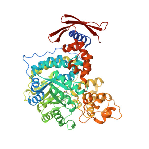
Biotin-dependent enzymes catalyze carboxyl transfer reactions by efficiently coordinating multiple reactions between spatially distinct active sites. Pyruvate carboxylase (PC), a multifunctional biotin-dependent enzyme, catalyzes the bicarbonate- and MgATP-dependent carboxylation of pyruvate to oxaloacetate, an important anaplerotic reaction in mammalian tissues. To complete the overall reaction, the tethered biotin prosthetic group must first gain access to the biotin carboxylase domain and become carboxylated and then translocate to the carboxyltransferase domain, where the carboxyl group is transferred from biotin to pyruvate. Here, we report structural and kinetic evidence for the formation of a substrate-induced biotin binding pocket in the carboxyltransferase domain of PC from Rhizobium etli. Structures of the carboxyltransferase domain reveal that R. etli PC occupies a symmetrical conformation in the absence of the biotin carboxylase domain and that the carboxyltransferase domain active site is conformationally rearranged upon pyruvate binding. This conformational change is stabilized by the interaction of the conserved residues Asp(590) and Tyr(628) and results in the formation of the biotin binding pocket. Site-directed mutations at these residues reduce the rate of biotin-dependent reactions but have no effect on the rate of biotin-independent oxaloacetate decarboxylation. Given the conservation with carboxyltransferase domains in oxaloacetate decarboxylase and transcarboxylase, the structure-based mechanism described for PC may be applicable to the larger family of biotin-dependent enzymes.
Organizational Affiliation:
Department of Biological Sciences, Marquette University, Milwaukee, Wisconsin 53201, USA.


