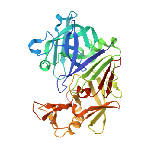
4KUP

Endothiapepsin in complex with 20mM acylhydrazone inhibitor


Experimental Data Snapshot

  • Method: X-RAY DIFFRACTION
  • Resolution: 1.31 Å
  • R-Value Free: 0.149 
  • R-Value Work: 0.130 
  • R-Value Observed: 0.131 

Starting Model: experimental
View more details

wwPDB Validation   3D Report Full Report


Ligand Structure Quality Assessment 


This is version 1.2 of the entry. See complete history


Literature

Structure-based design of inhibitors of the aspartic protease endothiapepsin by exploiting dynamic combinatorial chemistry.

Mondal, M.Radeva, N.Koster, H.Park, A.Potamitis, C.Zervou, M.Klebe, G.Hirsch, A.K.

(2014) Angew Chem Int Ed Engl 53: 3259-3263

  • DOI: https://doi.org/10.1002/anie.201309682
  • Primary Citation of Related Structures:  
    4KUP, 4LBT, 4LHH

  • PubMed Abstract: 

    Structure-based design (SBD) can be used for the design and/or optimization of new inhibitors for a biological target. Whereas de novo SBD is rarely used, most reports on SBD are dealing with the optimization of an initial hit. Dynamic combinatorial chemistry (DCC) has emerged as a powerful strategy to identify bioactive ligands given that it enables the target to direct the synthesis of its strongest binder. We have designed a library of potential inhibitors (acylhydrazones) generated from five aldehydes and five hydrazides and used DCC to identify the best binder(s). After addition of the aspartic protease endothiapepsin, we characterized the protein-bound library member(s) by saturation-transfer difference NMR spectroscopy. Cocrystallization experiments validated the predicted binding mode of the two most potent inhibitors, thus demonstrating that the combination of de novo SBD and DCC constitutes an efficient starting point for hit identification and optimization.


  • Organizational Affiliation

    Stratingh Institute for Chemistry, University of Groningen, Nijenborgh 7, 9747 AG Groningen (The Netherlands) http://www.rug.nl/research/bio-organic-chemistry/hirsch/


Macromolecules
Find similar proteins by:  (by identity cutoff)  |  3D Structure
Entity ID: 1
MoleculeChains Sequence LengthOrganismDetailsImage
Endothiapepsin330Cryphonectria parasiticaMutation(s): 0 
EC: 3.4.23.22
UniProt
Find proteins for P11838 (Cryphonectria parasitica)
Explore P11838 
Go to UniProtKB:  P11838
Entity Groups  
Sequence Clusters30% Identity50% Identity70% Identity90% Identity95% Identity100% Identity
UniProt GroupP11838
Sequence Annotations
Expand
  • Reference Sequence
Small Molecules
Ligands 4 Unique
IDChains Name / Formula / InChI Key2D Diagram3D Interactions
1TZ
Query on 1TZ

Download Ideal Coordinates CCD File 
B [auth A],
C [auth A]
(2S)-2-azanyl-3-(3H-indol-3-yl)-N-[(E)-(2,4,6-trimethylphenyl)methylideneamino]propanamide
C21 H24 N4 O
KKEDWXLWEPQNQM-OGBFJMLUSA-N
GOL
Query on GOL

Download Ideal Coordinates CCD File 
I [auth A],
J [auth A],
K [auth A],
L [auth A]
GLYCEROL
C3 H8 O3
PEDCQBHIVMGVHV-UHFFFAOYSA-N
DMS
Query on DMS

Download Ideal Coordinates CCD File 
D [auth A],
E [auth A],
F [auth A],
G [auth A],
H [auth A]
DIMETHYL SULFOXIDE
C2 H6 O S
IAZDPXIOMUYVGZ-UHFFFAOYSA-N
ACT
Query on ACT

Download Ideal Coordinates CCD File 
M [auth A]ACETATE ION
C2 H3 O2
QTBSBXVTEAMEQO-UHFFFAOYSA-M
Experimental Data & Validation

Experimental Data

  • Method: X-RAY DIFFRACTION
  • Resolution: 1.31 Å
  • R-Value Free: 0.149 
  • R-Value Work: 0.130 
  • R-Value Observed: 0.131 
  • Space Group: P 1 21 1
Unit Cell:
Length ( Å )Angle ( ˚ )
a = 45.406α = 90
b = 72.694β = 109.32
c = 52.819γ = 90
Software Package:
Software NamePurpose
MAR345dtbdata collection
PHASERphasing
PHENIXrefinement
HKL-2000data reduction
HKL-2000data scaling

Structure Validation

View Full Validation Report


Ligand Structure Quality Assessment 


Entry History 

Deposition Data

Revision History  (Full details and data files)

  • Version 1.0: 2014-04-02
    Type: Initial release
  • Version 1.1: 2023-09-20
    Changes: Data collection, Database references, Derived calculations, Refinement description
  • Version 1.2: 2024-11-06
    Changes: Structure summary